

Ring array transducers for real-time 3-D imaging of an atrial septal occluder
- PMID: 22698504
- PMCID: PMC3434960
- DOI: 10.1016/j.ultrasmedbio.2012.03.019
Ring array transducers for real-time 3-D imaging of an atrial septal occluder
Abstract
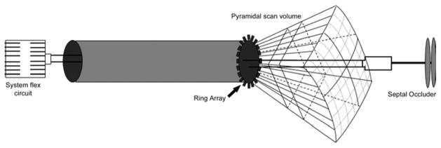
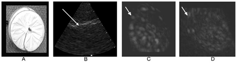
We developed new miniature ring array transducers integrated into interventional device catheters such as used to deploy atrial septal occluders. Each ring array consisted of 55 elements operating near 5 MHz with interelement spacing of 0.20 mm. It was constructed on a flat piece of copper-clad polyimide and then wrapped around an 11 French O.D. catheter. We used a braided cabling technology from Tyco Electronics Corporation to connect the elements to the Volumetric Medical Imaging (VMI) real-time 3-D ultrasound scanner. Transducer performance yielded a -6 dB fractional bandwidth of 20% centered at 4.7 MHz without a matching layer vs. average bandwidth of 60% centered at 4.4 MHz with a matching layer. Real-time 3-D rendered images of an en face view of a Gore Helex septal occluder in a water tank showed a finer texture of the device surface from the ring array with the matching layer.
Copyright © 2012 World Federation for Ultrasound in Medicine & Biology. Published by Elsevier Inc. All rights reserved.
Figures


Similar articles
-
New fabrication techniques for ring-array transducers for real-time 3D intravascular ultrasound.Ultrason Imaging. 2009 Oct;31(4):247-56. doi: 10.1177/016173460903100403. Ultrason Imaging. 2009. PMID: 20458877 Free PMC article.
-
A miniaturized catheter 2-D array for real-time, 3-D intracardiac echocardiography.IEEE Trans Ultrason Ferroelectr Freq Control. 2004 Oct;51(10):1334-46. doi: 10.1109/tuffc.2004.1350962. IEEE Trans Ultrason Ferroelectr Freq Control. 2004. PMID: 15553518
-
Double Ring Array Catheter for In Vivo Real-Time 3D Ultrasound.Ultrason Imaging. 2014 Jul;36(3):167-176. doi: 10.1177/0161734614523738. Epub 2014 Mar 12. Ultrason Imaging. 2014. PMID: 24626564
-
Real-time 3D laparoscopic ultrasonography.Ultrason Imaging. 2005 Jul;27(3):129-44. doi: 10.1177/016173460502700301. Ultrason Imaging. 2005. PMID: 16550704
-
Real-time 3-D ultrasound guidance of interventional devices.IEEE Trans Ultrason Ferroelectr Freq Control. 2008 Sep;55(9):2066-78. doi: 10.1109/TUFFC.898. IEEE Trans Ultrason Ferroelectr Freq Control. 2008. PMID: 18986903 Free PMC article.
Cited by
-
A Robotically Steerable Guidewire With Forward-Viewing Ultrasound: Development of Technology for Minimally-Invasive Imaging.IEEE Trans Biomed Eng. 2021 Jul;68(7):2222-2232. doi: 10.1109/TBME.2020.3042115. Epub 2021 Jun 17. IEEE Trans Biomed Eng. 2021. PMID: 33264091 Free PMC article.
References
-
- Degertekin FL, Guldiken RO, Karaman M. Annular-ring CMUT arrays for forward-looking IVUS: transducer characterization and imaging. IEEE Trans Ultrason Ferroelectr Freq Control. 2006;53:474–82. - PubMed
-
- Kaplan S. Congenital heart disease in adolescents and adults. Natural and postoperative history across age groups. Cardiol Clin. 1993;11:543–56. - PubMed
-
- Ko SF, Liang CD, Yip HK, Huang CC, Ng SH, Huang CF, Chen MC. Amplatzer septal occluder closure of atrial septal defect: evaluation of transthoracic echocardiography, cardiac CT, and transesophageal echocardiography. AJR Am J Roentgenol. 2009;193:1522–9. - PubMed
Publication types
MeSH terms
Grants and funding
LinkOut - more resources
Full Text Sources
Research Materials
Miscellaneous
