. 2012 Aug 15;368(2):382-92.
doi: 10.1016/j.ydbio.2012.06.008. Epub 2012 Jun 12.

Forward genetics uncovers Transmembrane protein 107 as a novel factor required for ciliogenesis and Sonic hedgehog signaling

Affiliations

Forward genetics uncovers Transmembrane protein 107 as a novel factor required for ciliogenesis and Sonic hedgehog signaling

Kasey J Christopher et al. Dev Biol. .

Abstract

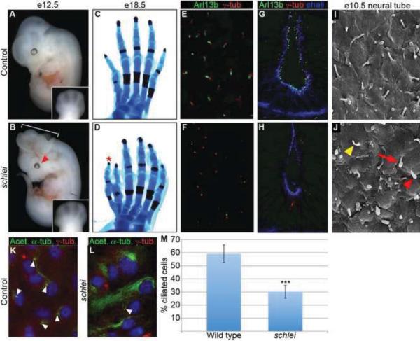
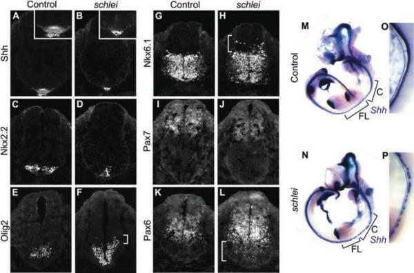
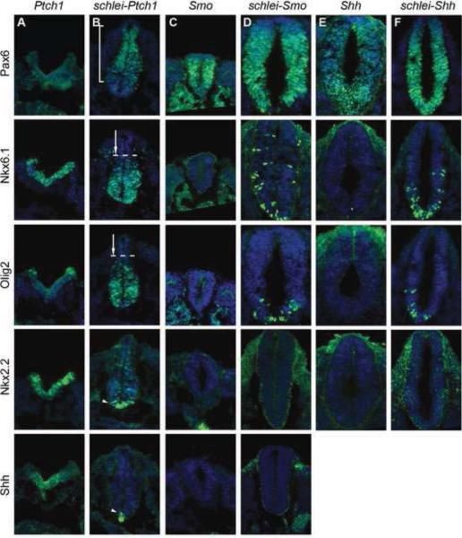
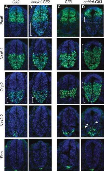
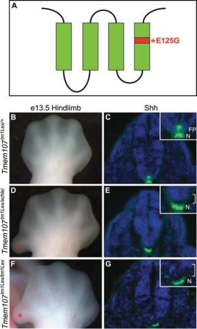
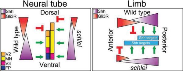
Cilia are dynamic organelles that are essential for a vast array of developmental patterning events, including left-right specification, skeletal formation, neural development, and organogenesis. Despite recent advances in understanding cilia form and function, many key ciliogenesis components have yet to be identified. By using a forward genetics approach, we isolated a novel mutant allele (schlei) of the mouse Transmembrane protein 107 (Tmem107) gene, which we show here is critical for cilia formation and embryonic patterning. Tmem107 is required for normal Sonic hedgehog (Shh) signaling in the neural tube and acts in combination with Gli2 and Gli3 to pattern ventral and intermediate neuronal cell types. schlei mutants also form extra digits, and we demonstrate that Tmem107 acts in the Shh pathway to determine digit number, but not identity, by regulating a subset of Shh target genes. Phenotypically, schlei mutants share several features with other cilia mutants; however, spatial restriction of mutant phenotypes and lack of left-right patterning defects in schlei animals suggest differential requirements for Tmem107 in cilia formation in distinct tissues. Also, in contrast to mutants with complete loss of cilia, schlei mutants retain some function of both Gli activator and repressor forms. Together, these studies identify a previously unknown regulator of ciliogenesis and provide insight into how ciliary factors affect Shh signaling and cilia biogenesis in distinct tissues.


Figures

Figure 1
Figure 1. schlei mutants display multiple developmental defects and reduced primary cilia formation
e12.5 schlei mutant embryos display broadening of the limbs (A,B) and a subset of mutants also show exencephaly (60%; n=140; bracket in B) or microphthalmia (66% at e12.5 and older; n=29; arrowhead in B). Early limb broadening resolves into preaxial polydactyly (asterisk) as shown in e18.5 hindlimbs (C,D). Confocal Z-stack projections of immunostaining of transverse cryosections through the limb (E,F) and neural tube (G,H) at e11.5 reveals a reduction in cilia number (as marked by Arl13b in green) in schlei animals (F,H) as compared to wild type (E,G) despite the normal appearance and localization of basal bodies (as marked by γ-tubulin in red). (I,J) Scanning electron microscopy confirms a reduction in number of cilia in the neural tube of schlei animals (J) as compared to wild type (I). Cilia that do form in schlei are malformed, including examples with bulges at the tip (red arrowhead), elongated and curled cilia (red arrow), or thin cilia (yellow arrowhead). Differentiated MEFs from e13.5 wild type (K) and schlei (L) embryos immunostained using acetylated α-tubulin reveal a reduction in cilia number in the mutants (cilia, arrowheads). (M) schlei mutant MEF lines showed fewer cilia than wild-type (n=4 lines of each examined). On average, 59.14 ± 6.75% of wild type cells formed cilia, compared to 30.22 ± 4.90% of mutant cells (p=0.00045 by two-tailed student's t-test with equal variances). In (C,D) hindlimbs are visualized with Alizarin red (bone) and Alcian blue (cartilage) staining. Blue staining in (G,H) = phalloidin. Blue staining in (K,L) = DAPI. Control and schlei mutant images are shown at the same magnification.
Figure 2
Figure 2. schlei mutants show disruptions in ventral cell type specification and neuronal subtype mixing in the neural tube
Sections through e10.5 neural tubes reveal that the floorplate is lost (A,B) and other ventral cell types including V3 interneuron progenitors (Nkx2.2+; C,D) are decreased in number and specified at more ventral locations than in wild type. Cells requiring intermediate levels of Shh signaling, such as motor neuron progenitors (Olig2+; E,F), are both specified more ventrally and expanded dorsally (bracket in F) in schlei. Other intermediate cell types, including a population marked by Nkx6.1 (G,H), are also expanded dorsally in the mutant and are mixed with more dorsal cell types (bracket in H). Some dorsal cell populations, such as the region demarcated by Pax7 expression, appear unchanged in schlei mutants (I,J). In contrast, Pax6 expression, which in wild type embryos (K) is inhibited by high levels of Shh, expands ventrally in schlei (bracket in L). The loss of the floorplate varies based on the axial level as visualized by whole mount in situ hybridization for Shh, with gaps in expression found particularly at the cervical and forelimb levels (M,N). (O,P) Higher magnification views of the cervical regions in e10.5 embryos highlight gaps in floorplate expression of Shh in schlei mutants. C; cervical; FL; forelimb. Control and schlei mutant images are shown at the same magnification.
Figure 3
Figure 3. The schlei mutation rescues ventral neuronal specification in Shh pathway mutants
e10.5 neural tubes stained for DAPI (blue) and neuronal markers (green). Expression of the dorsal marker Pax6, which is expanded in Shh (E) and Smo (C) mutants but absent in Ptch1 (A) is rescued and dorsally restricted in schlei-Ptch1 double mutants (B, bracket). In contrast, intermediate-level cells such as the Nkx6.1+ population and Olig2+ motor neuron progenitors, which require positive Shh signaling and are therefore absent in Shh and Smo, are able to form in schlei-Shh (F) and schlei-Smo (D) double mutants. These populations, which are expanded dorsally in Ptch1, are restored to a more ventral location in schlei-Ptch1 double mutants (arrows). High-level Shh targets (Nkx2.2+ V3 interneuron progenitors and Shh+ floorplate cells) are expanded dorsally in Ptch1 animals but are more ventrally restricted in schlei-Ptch1 double mutants (arrowheads). However, schlei is not able to rescue the loss of these cell types in Shh and Smo mutants. All neural tube sections are shown at the same magnification.
Figure 4
Figure 4. schlei interacts genetically with Gli3 and Gli2 to pattern the ventral and intermediate neural tube
e10.5 neural tubes stained for DAPI (blue) and neuronal markers (green). Expression of Pax6 is similar to wild type in Gli2 (A) and Gli3 (C) mutants, but the ventral border is extended ventrally in schlei-Gli2 (B, bracket), consistent with the schlei single mutant. In schlei-Gli3 embryos, there is a strong dorsal shift in the ventral border of Pax6 (D, arrow). The dorsal expansion of the Nkx6.1+ population observed in schlei is exacerbated in schlei-Gli3 (bracket) but rescued in schlei-Gli2. While intermediate cells such as Olig2+ motor neuron progenitors are comparable to wild type in Gli3 mutants, they are specified more ventrally in Gli2, schlei-Gli2, and schlei-Gli3 (brackets). Additionally, the dorsal boundary of expression of Olig2+ is pushed even further dorsally in schlei-Gli3 (upper bracket) than in schlei, but in schlei-Gli2 is restored to a level similar to wild type. Nkx2.2+ V3 progenitor cells are reduced in number and specified more ventrally in Gli2 mutants, but are completely lost in schlei-Gli2 double mutants. Nkx2.2+ cells are normal in Gli3, but are specified more ventrally and dorsally (arrowheads) in schlei-Gli3 double mutants. The floorplate, as marked by Shh, is normal in Gli3 but is absent in schlei-Gli3, Gli2, and schlei-Gli2 despite normal Shh staining in the notochord, similar to schlei mutants. All neural tube sections are shown at the same magnification.
Figure 5
Figure 5. Shh signaling appears expanded in schlei mutant limbs at e11.5
Despite a normal domain of Shh (A,B), the boundary (arrowhead) of expression of Gli1, a direct target of Shh, is expanded anteriorly in schlei mutants (C,D). Note also the increased width of the mutant limb relative to the control, an early indication of polydactyly (visible in B,D). Expression of Ptch1 appears unchanged in the mutant, suggesting differential sensitivities of various Shh targets (E,F). Gremlin, a downstream target of Shh activation, is expressed ectopically in a distinct region (arrow) in the anterior of the schlei limb (G,H). Control and schlei mutant images are shown at the same magnification.
Figure 6
Figure 6. The schlei mutation partially rescues digit number in Shh mutant limbs
Sox9 marks condensing digits in e13.5 forelimbs (Akiyama et al., 2002), revealing five digits in wild type (A), an ectopic anterior digit in schlei mutants (B), a single digit in Shh mutants (C), and at least four digits in the double mutant (D). The direct positive target of Shh, Gli1, is not activated in Shh (G) or the double mutant (H), compared with the anterior expansion of expression observed in schlei (F) relative to wild type (E). However, expression of Gremlin, which is anteriorly expanded in schlei (arrow in J) as compared to wild type (I), is partially restored in the double mutant (L) despite its complete absence in the Shh mutant (K), suggestive of a loss of Gli3R function. The rescue of digit number in schlei-Shh double mutants may be due in part to a rescue of cell death; TUNEL (green staining) in e10.5 forelimb sections demonstrates that, similar to wild type (M) and schlei (N), double mutant limbs (P) lack the extensive distal cell death observed in Shh mutants (O). Hoxd11 is expressed in the presumptive digits two through five in wild type (Q), and the ectopic digit in schlei variably expresses Hoxd11, indicating posterior identity in the example shown (R). Hoxd11 is absent in both the Shh mutant (S) and the double mutant (T). Blue staining in M–P = DAPI. Control and schlei mutant images are shown at the same magnification.
Figure 7
Figure 7. The schlei mutant phenotype results from a point mutation in Tmem107
(A) Schematic representation of Tmem107 protein, which is predicted to contain four transmembrane domains. Asterisk and red shading, location of the E125G missense mutation in schlei allele. Complementation analysis was carried out by crossing Tmem107tm1Lex males with schlei females. At e10.5, transheterozygous embryos demonstrated that Tmem107tm1Lex fails to complement schlei in anteroposterior limb patterning, as shown by preaxial polydactyly (asterisk) visible at e13.5 (B,D) and floorplate (FP) formation, as shown by staining for Shh (C,E). These phenotypes are also recapitulated in embryos homozygous for the Tmem107tm1Lex mutant allele (F,G). Blue staining in (C,E,G) = DAPI. N = notochord. Brackets in E,G indicate loss of floorplate. Control, transheterozygotes, and homozygous mutant images are shown at the same magnification.
Figure 8
Figure 8. Model of Gli activity in the schlei mutant
In the neural tube, a ventral gradient of Shh signaling specifies ventral neuronal cell types, while dorsal Gli3R helps restrict the extent of their domain. In the schlei ventral neural tube, the highest level of Shh responsiveness is lost, but there is an increased range of intermediate-level signaling extending more dorsally, mimicking a shallower, but elongated gradient. Gli3 repressor function is also diminished in schlei, and together, this results in the absence of the highest-level ventral target, the floorplate (FP), combined with a broadened domain competent to form motor neurons (MN) and V2 interneurons. In the limb, a posterior gradient of Shh signaling specifies digit identity while anterior Gli3R restricts digit number. Similar to the neural tube, the Shh gradient appears extended in schlei limbs and together with decreased Gli3R function results in an expansion of Shh target gene expression (Gremlin, Gli1) in the anterior portion of the limb.

Similar articles

Cited by

References

    1. Akiyama H, Chaboissier MC, Martin JF, Schedl A, de Crombrugghe B. The transcription factor Sox9 has essential roles in successive steps of the chondrocyte differentiation pathway and is required for expression of Sox5 and Sox6. Genes Dev. 2002;16(21):2813–28. - PMC - PubMed
    1. Ansley SJ, Badano JL, Blacque OE, Hill J, Hoskins BE, Leitch CC, Kim JC, Ross AJ, Eichers ER, Teslovich TM, et al. Basal body dysfunction is a likely cause of pleiotropic Bardet-Biedl syndrome. Nature. 2003;425(6958):628–33. - PubMed
    1. Avidor-Reiss T, Maer AM, Koundakjian E, Polyanovsky A, Keil T, Subramaniam S, Zuker CS. Decoding cilia function: defining specialized genes required for compartmentalized cilia biogenesis. Cell. 2004;117(4):527–39. - PubMed
    1. Bai CB, Auerbach W, Lee JS, Stephen D, Joyner AL. Gli2, but not Gli1, is required for initial Shh signaling and ectopic activation of the Shh pathway. Development. 2002;129(20):4753–61. - PubMed
    1. Bai CB, Joyner AL. Gli1 can rescue the in vivo function of Gli2. Development. 2001;128(24):5161–72. - PubMed

MeSH terms