

Hippocampal tissue of patients with refractory temporal lobe epilepsy is associated with astrocyte activation, inflammation, and altered expression of channels and receptors
- PMID: 22698689
- PMCID: PMC3412889
- DOI: 10.1016/j.neuroscience.2012.06.002
Hippocampal tissue of patients with refractory temporal lobe epilepsy is associated with astrocyte activation, inflammation, and altered expression of channels and receptors
Abstract
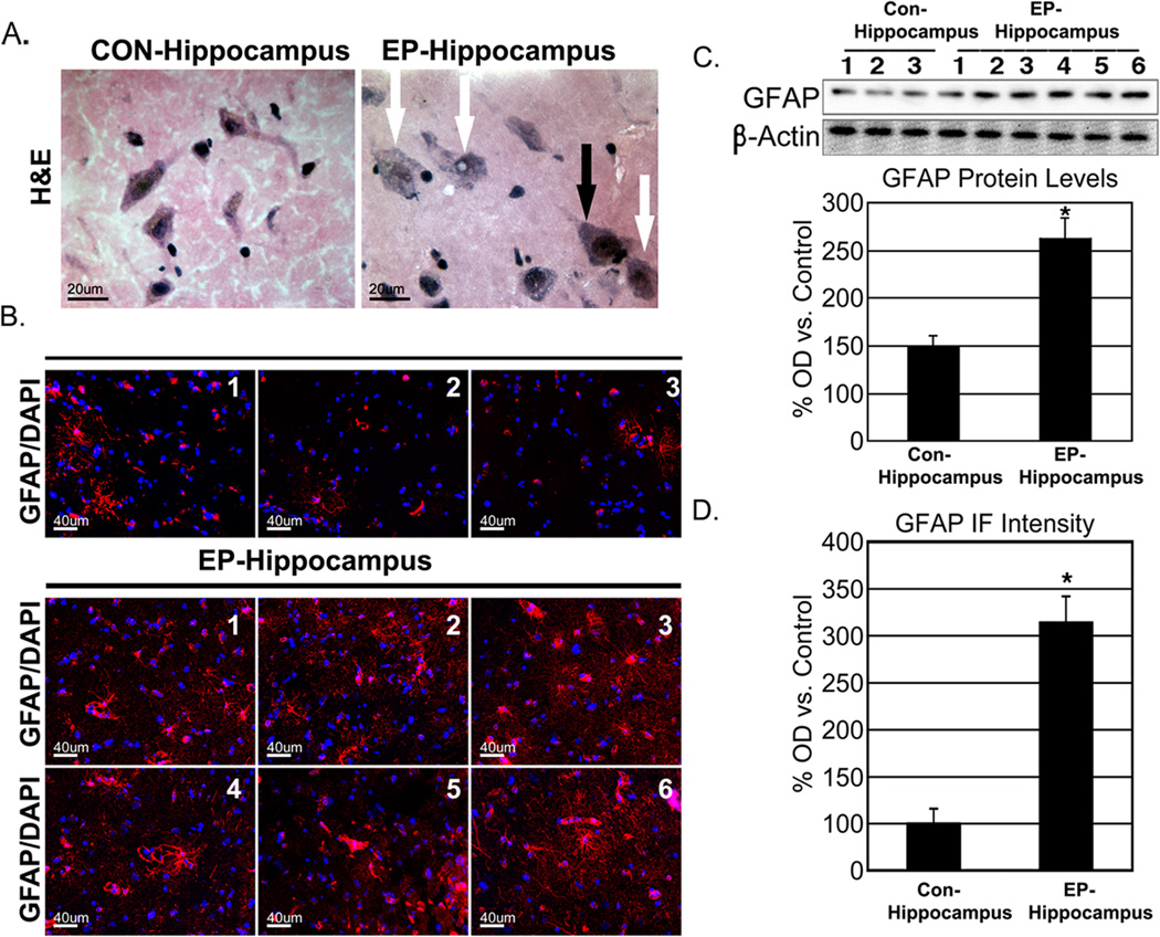
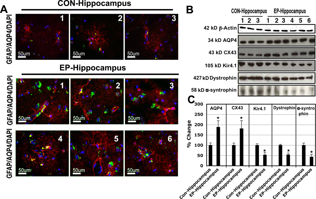
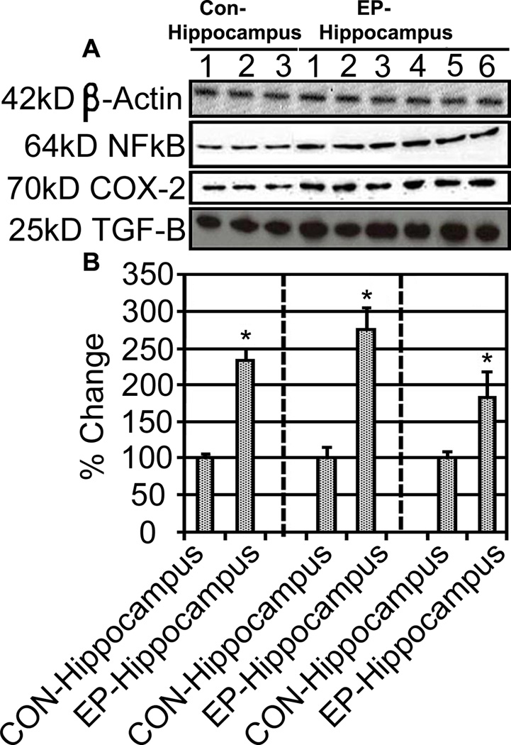
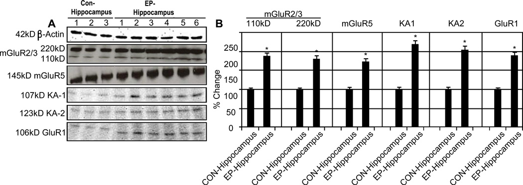
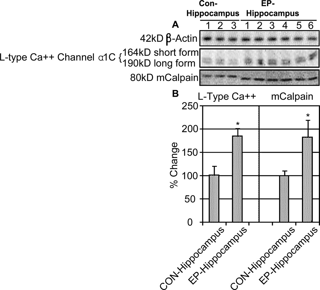
Temporal lobe epilepsy (TLE) is the most common form of focal epilepsy. Previous research has demonstrated several trends in human tissue that, undoubtedly, contribute to the development and progression of TLE. In this study we examined resected human hippocampus tissue for a variety of changes including gliosis that might contribute to the development and presentation of TLE. The study subjects consisted of six TLE patients and three sudden-death controls. Clinicopathological characteristics were evaluated by H&E staining. Immunohistological staining and Western blotting methods were used to analyze the samples. Neuronal hypertrophy was observed in resected epileptic tissue. Immunohistological staining demonstrated that activation of astrocytes was significantly increased in epileptic tissue as compared to corresponding regions of the control group. The Western blot data also showed increased CX43 and AQP4 in the hippocampus and downregulation of Kir4.1, α-syntrophin, and dystrophin, the key constituents of AQP4 multi-molecular complex. These tissues also demonstrated changes in inflammatory factors (COX-2, TGF-β, NF-κB) suggesting that these molecules may play an important role in TLE pathogenesis. In addition we detected increases in metabotropic glutamate receptor (mGluR) 2/3, mGluR5 and kainic acid receptor subunits KA1 (Grik4) and KA2 (Grik5) in patients' hippocampi. We noted increased expression of the α1c subunit comprising class C L-type Ca(2+) channels and calpain expression in these tissues, suggesting that these subunits might have an integral role in TLE pathogenesis. These changes found in the resected tissue suggest that they may contribute to TLE and that the kainic acid receptor (KAR) and deregulation of GluR2 receptor may play an important role in TLE development and disease course. This study identifies alterations in number of commonly studied molecular targets associated with astrogliosis, cellular hypertrophy, water homeostasis, inflammation, and modulation of excitatory neurotransmission in hippocampal tissues from TLE patients.
Published by Elsevier Ltd.
Conflict of interest statement
None of the authors has any conflict of interest to disclose. We confirm that we have read the Journal’s position on issues involved in ethical publication and affirm that this report is consistent.
Figures
References
-
- Araújo IM, Gil JM, Carreira BP, Mohapel P, Petersen A, Pinheiro PS, Soulet D, Bahr BA, Brundin P, Carvalho CM. Calpain activation is involved in early caspase-independent neurodegeneration in the hippocampus following status epilepticus. J Neurochem. 2008;105:666–676. - PubMed
-
- Aronica E, van Vliet EA, Mayboroda OA, Troost D, da Silva FH, Gorter JA. Upregulation of metabotropic glutamate receptor subtype mGluR3 and mGluR5 in reactive astrocytes in a rat model of mesial temporal lobe epilepsy. Eur J Neurosci. 2000;12:2333–2344. - PubMed
-
- Benarroch EE. Astrocyte-neuron interactions: implications for epilepsy. Neurology. 2009;73:1323–1327. - PubMed
-
- Broberg M, Pope KJ, Lewis T, Olsson T, Nilsson M, Willoughby JO, et al. Cell swelling precedes seizures induced by inhibition of astrocytic metabolism. Epilepsy Res. 2008;80:132–141. - PubMed
Publication types
MeSH terms
Grants and funding
LinkOut - more resources
Full Text Sources
Research Materials
Miscellaneous
