. 2012 Oct;61(10):2484-94.
doi: 10.2337/db11-1665. Epub 2012 Jun 14.

Metformin inhibits growth hormone-mediated hepatic PDK4 gene expression through induction of orphan nuclear receptor small heterodimer partner

Affiliations

Metformin inhibits growth hormone-mediated hepatic PDK4 gene expression through induction of orphan nuclear receptor small heterodimer partner

Yong Deuk Kim et al. Diabetes. 2012 Oct.

Abstract

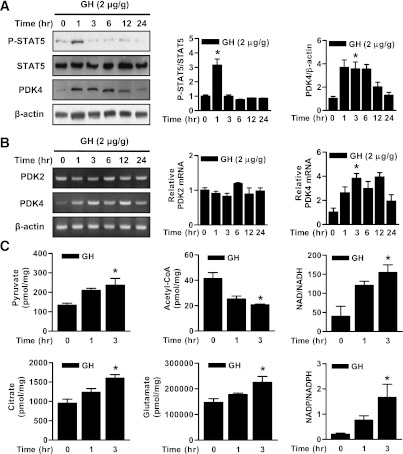
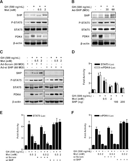
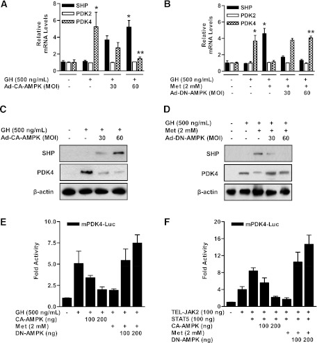
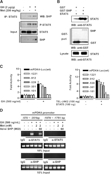
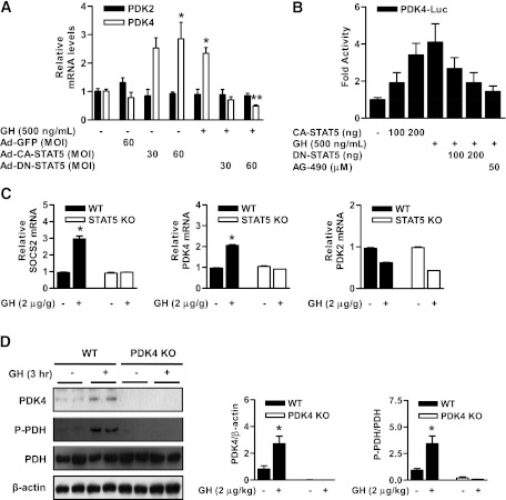
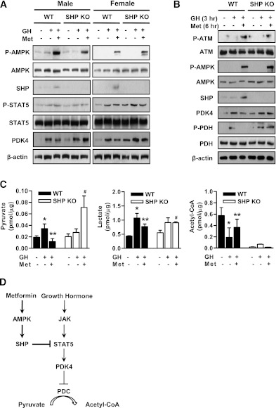
Growth hormone (GH) is a counter-regulatory hormone that plays an important role in preventing hypoglycemia during fasting. Because inhibition of the pyruvate dehydrogenase complex (PDC) by pyruvate dehydrogenase kinase 4 (PDK4) conserves substrates for gluconeogenesis, we tested whether GH increases PDK4 expression in liver by a signaling pathway sensitive to inhibition by metformin. The effects of GH and metformin were determined in the liver of wild-type, small heterodimer partner (SHP)-, PDK4-, and signal transducer and activator of transcription 5 (STAT5)-null mice. Administration of GH in vivo increased PDK4 expression via a pathway dependent on STAT5 phosphorylation. Metformin inhibited the induction of PDK4 expression by GH via a pathway dependent on AMP-activated protein kinase (AMPK) and SHP induction. The increase in PDK4 expression and PDC phosphorylation by GH was reduced in STAT5-null mice. Metformin decreased GH-mediated induction of PDK4 expression and metabolites in wild-type but not in SHP-null mice. In primary hepatocytes, dominant-negative mutant-AMPK and SHP knockdown prevented the inhibitory effect of metformin on GH-stimulated PDK4 expression. SHP directly inhibited STAT5 association on the PDK4 gene promoter. Metformin inhibits GH-induced PDK4 expression and metabolites via an AMPK-SHP-dependent pathway. The metformin-AMPK-SHP network may provide a novel therapeutic approach for the treatment of hepatic metabolic disorders induced by the GH-mediated pathway.


Figures

FIG. 1.
FIG. 1.
GH regulates PDK4 gene expression and liver metabolites. A: Wild-type mice (12 weeks old) were injected i.p. with GH (2 μg/g body weight) at the observed time periods. Tissue extracts prepared from the tissues of the indicated groups were subjected to Western blot analysis with various antibodies. Protein levels were normalized to total form antibodies and/or β-actin levels. All mice were separated into experimental groups (n = 4–6 mice/group). *P < 0.05 compared with untreated controls. B: WT mice were determined at the indicated time periods after the administration of GH in the feeding condition. Total RNA extracts were isolated from liver samples at the indicated time periods after GH treatment. The levels of PDK2 and PDK4 mRNA were measured by RT-PCR analysis and then normalized to internal control (β-actin level). *P < 0.05 compared with untreated controls. C: Liver samples were measured at the observed time periods after GH treatment. Metabolites were analyzed with the liquid chromatography-mass spectrometry/mass spectrometry method as described in research design and methods. All mice were separated into experimental groups (n = 4 mice/group). *P < 0.05 compared with untreated controls.
FIG. 2.
FIG. 2.
Metformin inhibits GH-induced STAT5 transactivity and PDK4 expression in primary hepatocytes. Rat primary hepatocytes were cultured under serum-free conditions for 24 h. A: After pretreatment with metformin (Met) for 12 h, the cells were treated with GH for 1 h at the indicated dose (500 ng/mL). B: Rat primary hepatocytes were infected with Ad-SHP at a multiplicity of infection (MOI) of 30 or 60 for 36 h. After infection for 36 h, cells were treated with GH (500 ng/mL) for 1 h. Specific proteins were assayed in whole-cell extracts by Western blot analysis with the indicated antibodies and then normalized to an internal control (β-actin level and/or total forms). C: Rat primary hepatocytes were infected with 60 MOI of Ad-siSHP and Ad-Scram for 36 h. After infection with Ad-siSHP and Ad-Scram, cells were treated with GH (500 ng/mL) for 1 h, with or without metformin for 12 h at the indicated dose. Whole-cell extracts were isolated and analyzed by immunoblotting with the indicated antibodies, and then normalized to an internal control (β-actin level and/or total forms). The results shown are representative of at least three independent experiments. D: HepG2 cell lines were cotransfected with SHP in the indicated reporter genes and treated with GH for 1 h or metformin for 12 h after transfection. Luciferase (Luc) activity was measured after 36 h and normalized to β-galactosidase activity. All data are representative of three independently performed experiments and are shown as fold activations relative to the control (± SEM). E and F: HepG2 cell lines were transfected with the oligonucleotide siSHP and siScram. After transfection for 36 h, cells were transfected with the indicated reporter gene (STAT5-Luc [E], mPDK4-Luc [F]) and then treated with GH (500 ng/mL) for 1 h in the presence of metformin for 12 h. Luciferase (Luc) activity was normalized to β-galactosidase activity. The results shown are representative of at least three independent experiments. All data are indicated as fold activations relative to the control (± SEM).
FIG. 3.
FIG. 3.
Induction of SHP by AMPK inhibits GH-induced PDK4 expression in primary hepatocytes. A: Rat primary hepatocytes were infected with 30, 60 multiplicity of infection (MOI) of adenoviral vector expressing CA-AMPK for 36 h. After infection with Ad-CA-AMPK, cells were treated with GH (500 ng/mL) for 1 h. Total RNA extracts were isolated and analyzed by RT-PCR analysis with the indicated primers and then normalized to an internal control (β-actin level). *P < 0.05 and **P < 0.05 compared with untreated control, GH-treated cells. B: Rat primary hepatocytes were infected with Ad-DN-AMPK for 36 h and then treated with GH (500 ng/mL) for 1 h in the presence or absence of metformin (Met) for 12 h. Total RNA were isolated from hepatocytes and used by RT-PCR analysis. SHP, PDK2, and PDK4 mRNA levels were normalized to an internal control with β-actin level. *P < 0.05 and **P < 0.01 compared with untreated control, GH-treated cells. Rat primary hepatocytes were infected with Ad-CA-AMPK (C) and Ad-DN-AMPK (D) for 30 or 60 MOI for 36 h. After infection, cells were treated with GH (500 ng/mL) for 1 h with or without metformin for 12 h. Specific proteins were determined by Western blot analysis with the indicated antibodies, and then normalized to an internal control (β-actin level and/or total forms). E: HepG2 cells were cotransfected with CA-AMPK and DN-AMPK and then treated with GH (500 ng/mL) for 1 h in the presence of metformin for 12 h. F: Cells were cotransfected with TEL-JAK2, STAT5, CA-AMPK, and DN-AMPK and then treated with metformin for 12 h. Luciferase (Luc) activity was normalized to β-galactosidase activity to correct for variations in transfection efficiency. All data are representative of at least three independent experiments. All data are shown as fold activations relative to the control (± SEM).
FIG. 4.
FIG. 4.
Interaction of between SHP and STAT5 in vivo. A: Wild-type mice were injected with GH (2 μg/g body weight) for 1 h or metformin (Met; 200 mg/kg body weight) for 6 h at the indicated concentrations. Coimmunoprecipitation assays with liver extracts demonstrate the functional association between SHP and STAT5. Tissue extracts (700 μg/lane) were immunoprecipitated with STAT5 antibody and blotted with SHP antibody. Expression of p-STAT5, STAT5, and SHP from 10% input were analyzed by immunoblotting. All mice were separated into experimental groups (n = 4–6 mice/group). B: In vivo interaction between SHP and STAT5. HepG2 cells were cotransfected with expression vectors for STAT5 together with p-EBG-SHP (GST-SHP) and p-EBG (GST alone) as a control. The complex formation (top) and the amount of STAT5 used for the in vivo binding assay (bottom, lysate) were analyzed by Western blot (WB) using anti-STAT5 antibody. The same blot was stripped and reprobed with an anti-GST antibody (middle) to confirm the expression levels of the GST-SHP and the GST control. C: Schematic diagrams show the wild-type (wt) and the deletion forms of the mPDK4 promoter constructs. HepG2 cells were cotransfected with wild-type, deletion form mPDK4 reporter, and TEL-JAK2, STAT5, respectively. After transfection for 36 h, cells were treated with GH (500 ng/mL) for 1 h. Luciferase (Luc) activity was normalized to β-galactosidase activity to correct for transfection efficiency. All data are shown as fold activations relative the control (± SEM). D: ChIP assay. Rat primary hepatocytes were infected with Ad-siSHP for 36 h and then treated with GH and metformin at the indicated concentrations. Before immunoprecipitation, an aliquot of the sample was stored and then purified and represents input for each sample. Cell extracts were immunoprecipitated with STAT5 and SHP antibodies, and purified DNA samples were used to perform PCR using primers binding the specific proximal (left) and nonspecific distal (right) regions on the mPDK4 gene promoter. All data are representative of at least three independent experiments.
FIG. 5.
FIG. 5.
GH-mediated induction of PDC phosphorylation is mediated by the STAT5-PDK4 pathway in the liver. A: Rat primary hepatocytes were infected with Ad-GFP, Ad-CA-STAT5, and Ad-DN-STAT5 for 36 h and then treated with GH for 3 h. Total RNA extracts were isolated and analyzed by RT-PCR analysis with the indicated primers and then normalized to an internal control (β-actin level). *P < 0.05 and **P < 0.05 compared with untreated control, GH-treated cells. B: HepG2 cells were cotransfected with CA-STAT5 and DN-STAT5 at the indicated reporter gene and then treated with GH for 3 h or the JAK2 inhibitor AG490 for 6 h after transfection for 36 h. Luciferase (Luc) activity was normalized to β-galactosidase activity to correct for variations in transfection efficiency. All data are shown as fold activations relative to the control (± SEM). C: For GH stimulation, mice were injected with GH (2 μg/g body weight i.p.), killed 4 h after injection, and livers were harvested for analyses. Total RNA was isolated from liver tissues of Stat5f/f (wild-type [WT]) and Stat5f/f;Alb-Cre (liver-specific STAT5 KO) mice. The levels of PDK2, PDK4, and SOCS2 mRNA were measured by RT-PCR analysis and then normalized to internal control (β-actin level). Three mice from each experimental group were evaluated. *P < 0.05 compared with untreated wild-type mice. D: WT and PDK4-null mice were injected with GH (2 μg/g body weight i.p.) for 3 h. Tissue extracts were isolated from livers of the indicated groups and assessed by Western blot analysis with various antibodies. The protein levels were normalized to total form antibody and/or β-actin level. All mice were separated into experimental groups (n = 5 mice/group). *P < 0.05 compared with untreated wild-type mice.
FIG. 6.
FIG. 6.
Metformin (Met) alters GH-dependent pathway and liver metabolites through SHP induction in vivo. A: Male and female wild-type (WT) and SHP-null mice were orally injected with metformin (200 mg/kg body weight) for 6 h and then intraperitoneally treated with GH (2 μg/g body weight) for 1 h in the feeding condition. Tissue extracts were isolated from liver tissue harvested from the mice of the indicated groups and assessed by Western blot analysis with appropriate antibodies. Protein levels were normalized to total form antibodies and/or β-actin levels. All mice were separated into experimental groups (n = 4–6 mice/group). B: WT and SHP-null mice were orally administered with metformin (200 mg/kg body weight) for 6 h and then intraperitoneally treated with GH (2 μg/g body weight) for 3 h in the fed condition. Tissue extracts were isolated from liver tissue harvested from the mice of the indicated groups and assessed by Western blot analysis with appropriate antibodies. Protein levels were normalized to total form and/or β-actin levels. All mice were separated into experimental groups (n = 4–6 mice/group). C: WT and SHP-null mice were measured at the observed time periods after metformin and GH treatment. Metabolites were analyzed with the liquid chromatography-mass spectrometry/mass spectrometry method as described in research design and methods. *P < 0.05, **P < 0.005, and #P < 0.005 compared with untreated control wild-type mice, GH-treated wild-type mice, and GH- and Met-treated wild-type mice. D: Growth hormone upregulates PDK4 gene expression by STAT5 transactivation, whereas metformin, a known activator of SHP via AMPK pathway, represses the GH-STAT5 pathway via downregulation of DNA-binding activity of STAT5 on the PDK4 gene promoter.

Similar articles

Cited by

References

    1. Zhou G, Myers R, Li Y, et al. Role of AMP-activated protein kinase in mechanism of metformin action. J Clin Invest 2001;108:1167–1174 - PMC - PubMed
    1. Hundal RS, Krssak M, Dufour S, et al. Mechanism by which metformin reduces glucose production in type 2 diabetes. Diabetes 2000;49:2063–2069 - PMC - PubMed
    1. White MF. Metformin and insulin meet in a most atypical way. Cell Metab 2009;9:485–487 - PubMed
    1. Edgerton DS, Johnson KM, Cherrington AD. Current strategies for the inhibition of hepatic glucose production in type 2 diabetes. Front Biosci 2009;14:1169–1181 - PubMed
    1. Zhang BB, Zhou G, Li C. AMPK: an emerging drug target for diabetes and the metabolic syndrome. Cell Metab 2009;9:407–416 - PubMed

Publication types

MeSH terms

Substances