

Novel in vitro model for studying hepatic ischemia-reperfusion injury using liver cubes
- PMID: 22698934
- PMCID: PMC4258692
- DOI: 10.1016/j.surg.2012.02.012
Novel in vitro model for studying hepatic ischemia-reperfusion injury using liver cubes
Abstract
Background: Although inflow occlusion techniques have given surgeons the ability to carry out increasingly complex liver resections, ischemia-reperfusion (IR) injury continues to be a source of morbidity. Efforts to ameliorate IR injury have been hindered in absence of adequate preclinical models. The goal of the present study was to develop a simple, efficient, and cost-effective means of studying hepatic IR injury.
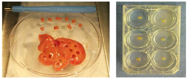
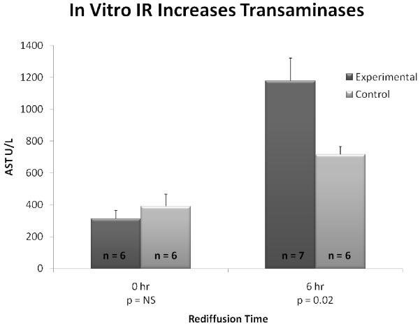
Methods: Liver cubes were procured from normal (C57BL/6) mice. After hepatectomy, 4-mm punch biopsies were taken for individual placement in culture wells containing hepatocyte media. Experimental cubes underwent hypoxia for 60 minutes, whereas controls remained normoxic. Supernatants were collected from individual wells after 0, 6, and 12 hours of rediffusion for transaminase and cytokine measurement. Histologic examination was performed on individual cubes.
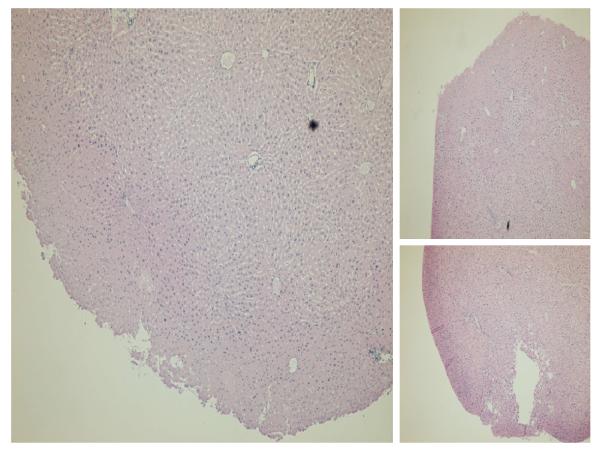
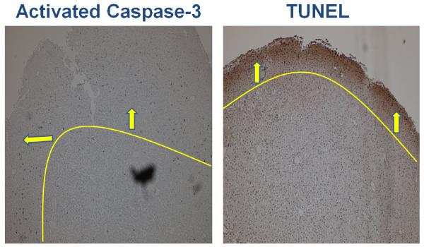
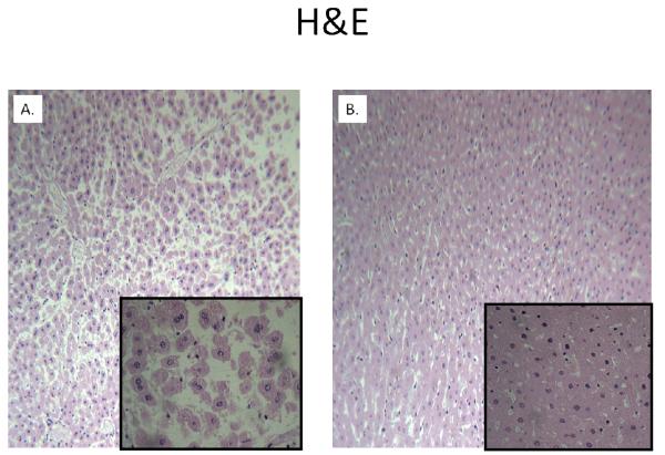
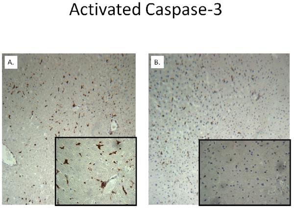
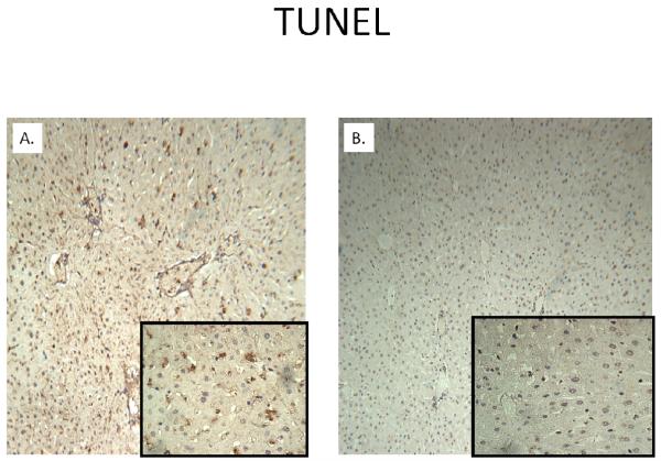
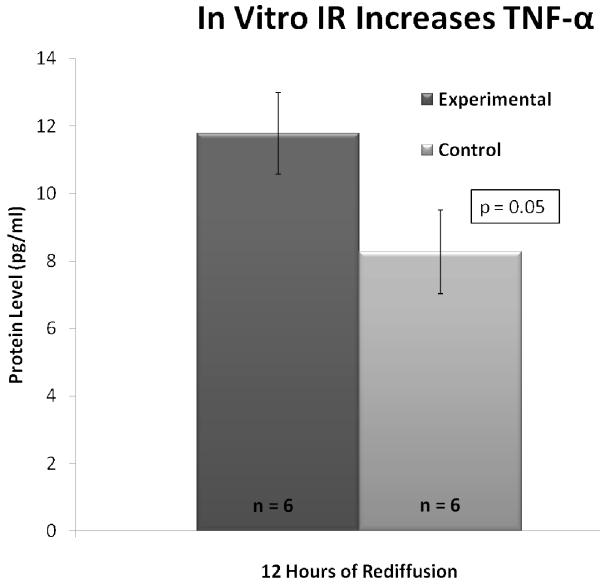
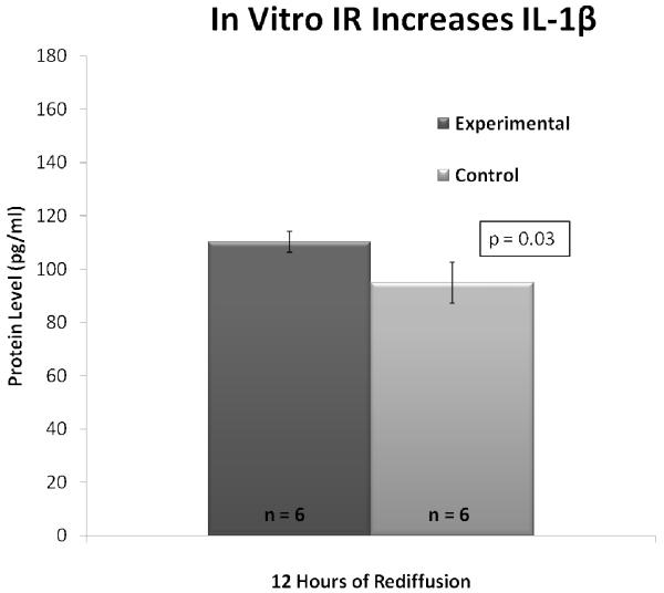
Results: Extensive histologic injury was seen in the experimental cubes compared with controls with greater staining for activated caspase-3 and terminal deoxynucleotidyl transferase dUTP nick end labeling at 6 and 24 hours, respectively. Changes consistent with ischemic injury occurred more centrally in liver cubes, whereas markers for rediffusion injury were appreciated along the periphery. Transaminases were significantly higher at 6 hours after rediffusion in experimental cubes compared with controls (P = .02). tumor necrosis factor-α and interleukin-1β were significantly higher in the media of experimental cubes compared with controls at 12 hours rediffusion (P = .05 and .03 respectively).
Conclusion: In vitro IR of cubes produces a significant injury with a pattern reflective of hepatic lobular architecture. This novel technique may open new avenues for uncoupling the mechanisms of IR while facilitating rapid screening of potential therapies.
Copyright © 2012 Mosby, Inc. All rights reserved.
Figures
Similar articles
-
Ischemic preconditioning improves liver tolerance to congestion-reperfusion injury in mice.J Surg Res. 2014 Jun 1;189(1):152-8. doi: 10.1016/j.jss.2014.01.061. Epub 2014 Feb 5. J Surg Res. 2014. PMID: 24589179
-
Reduction of hepatic ischemia/reperfusion injury by a soluble P-selectin glycoprotein ligand-1.Ann Surg. 1998 Jun;227(6):832-40. doi: 10.1097/00000658-199806000-00006. Ann Surg. 1998. PMID: 9637546 Free PMC article.
-
GGsTop, a novel and specific γ-glutamyl transpeptidase inhibitor, protects hepatic ischemia-reperfusion injury in rats.Am J Physiol Gastrointest Liver Physiol. 2016 Aug 1;311(2):G305-12. doi: 10.1152/ajpgi.00439.2015. Epub 2016 Jun 30. Am J Physiol Gastrointest Liver Physiol. 2016. PMID: 27365338
-
Cathepsin B inactivation attenuates the apoptotic injury induced by ischemia/reperfusion of mouse liver.Apoptosis. 2005 Dec;10(6):1261-9. doi: 10.1007/s10495-005-2358-1. Apoptosis. 2005. PMID: 16215674
-
Proliferative activity in ischemia/reperfusion injury in hepatectomized mice: effect of N-acetylcysteine.Transplant Proc. 2012 Oct;44(8):2321-5. doi: 10.1016/j.transproceed.2012.07.009. Transplant Proc. 2012. PMID: 23026584
Cited by
-
In Vitro Models for the Study of Liver Biology and Diseases: Advances and Limitations.Cell Mol Gastroenterol Hepatol. 2023;15(3):559-571. doi: 10.1016/j.jcmgh.2022.11.008. Epub 2022 Nov 26. Cell Mol Gastroenterol Hepatol. 2023. PMID: 36442812 Free PMC article. Review.
References
-
- Klune JR, Tsung A. Molecular biology of liver ischemia/reperfusion injury: established mechanisms and recent advancements. Surg Clin North Am. 2010;90(4):665–77. - PubMed
-
- Brancatisano R, Isla A, Habib N. Is radical hepatic surgery safe? Am J Surg. 1998;175(2):161–3. - PubMed
-
- Wanner GA, Ertel W, Muller P, Hofer Y, Leiderer R, Menger MD, et al. Liver ischemia and reperfusion induces a systemic inflammatory response through Kupffer cell activation. Shock. 1996;5(1):34–40. - PubMed
-
- Matuschak GM, Rinaldo JE. Organ interactions in the adult respiratory distress syndrome during sepsis. Role of the liver in host defense. Chest. 1988;94(2):400–6. - PubMed
-
- Kanoria S, Glantzounis G, Jalan R, Davies NA, Seifalian AM, Williams R, et al. A model to study total hepatic ischemia-reperfusion injury. Transplant Proc. 2004;36(9):2586–9. - PubMed
Publication types
MeSH terms
Substances
Grants and funding
LinkOut - more resources
Full Text Sources
Medical
Research Materials
