. 2012 Aug 20;162(1):1-8.
doi: 10.1016/j.jconrel.2012.06.006. Epub 2012 Jun 12.

Solid formulation of cell-penetrating peptide nanocomplexes with siRNA and their stability in simulated gastric conditions

Affiliations

Solid formulation of cell-penetrating peptide nanocomplexes with siRNA and their stability in simulated gastric conditions

Kariem Ezzat et al. J Control Release. .

Abstract

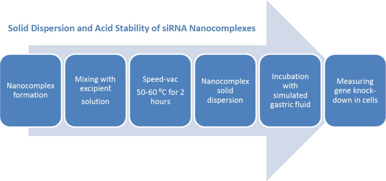
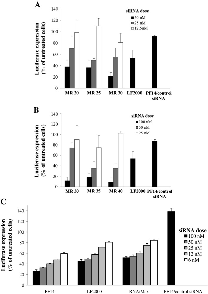
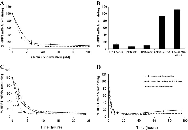
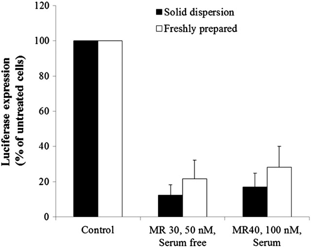
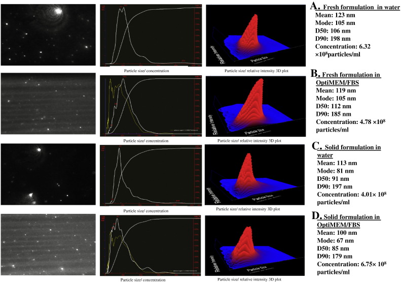
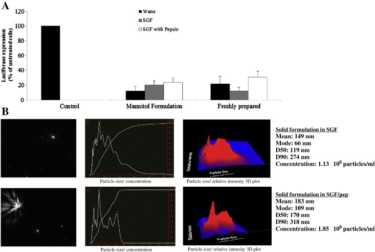
Cell-penetrating peptides (CPPs) are short cationic peptides that have been extensively studied as drug delivery vehicles for proteins, nucleic acids and nanoparticles. However, the formulation of CPP-based therapeutics into different pharmaceutical formulations and their stability in relevant biological environments have not been given the same attention. Here, we show that a newly developed CPP, PepFect 14 (PF14), forms non-covalent nanocomplexes with short interfering RNA (siRNA), which are able to elicit efficient RNA-interference (RNAi) response in different cell-lines. RNAi effect is obtained at low siRNA doses with a unique kinetic profile. Furthermore, the solid dispersion technique is utilized to formulate PF14/siRNA nanocomplexes into solid formulations that are as active as the freshly prepared nanocomplexes in solution. Importantly, the nanocomplexes are stable and active in mediating RNAi response after incubation with simulated gastric fluid (SGF) that is highly acidic. These results demonstrate the activity of PF14 in delivering and protecting siRNA in different pharmaceutical forms and biological environments.


Figures

None
Graphical abstract
Fig. 1
Fig. 1
PF14 mediates efficient RNAi in reporter cell-lines. BHK-21 cells and HepG2 (5 × 104) were seeded 24 h prior to experiments into 24-well plates. Cells were treated with PF14/siRNA nanocomplexes at three different concentrations at MRs 20:1, 25:1 and 30:1 for 4 h in serum-free medium followed by addition of serum to final concentration of 10% and incubated additionally for 20 h (A), and at MRs 30:1, 35:1 and 40:1 for 24 h in serum-containing medium (B). (C) For HepG2 cell-line, the cells were treated with PF14/siRNA nanocomplexes at different concentrations at MR30 in serum-containing medium. PF14 complexed with control (unrelated) siRNA was used at the highest siRNA dose and the highest MR in A, B and C. LF 2000® and LF RNAiMax® were used according to the manufacturer's protocol. Cells were lysed in 0.1% Triton X-100 and luciferase activity was measured. RNAi assay results are presented as percent of luciferase expression relative to untreated cells. The values represent the mean of at least three experiments performed in duplicate (mean ± SEM, n = 3).
Fig. 2
Fig. 2
PF14 induces knock-down of an endogenous gene with a unique kinetic profile. A. Dose–response curves for the down-regulation of HPRT1 by PF14/siRNA nanocomplexes in HUH7 cells in serum-containing medium (solid line) and in serum-free medium (dotted line). The concentration at which 50% of HPRT1 mRNA was depleted (EC50) was 12 nM and 8 nM for the serum and serum-free conditions respectively. B. Comparison between the efficiency of one selected dose (50 nM) of siRNA transfected by PF14 or by LF RNAimax® or naked siRNA without any transfection reagent. Control siRNA of unrelated sequence at the same concentration transfected with PF14 was also included. C and D: Kinetics of the HPRT1 down-regulation using a 50 nM dose of siRNA transfected with PF14 at serum-containing and serum-free conditions and with LF RNAimax®. C. represents the kinetics up to 24 h while D. shows the time-course study as monitored up to 4 days. Significant differences were detected in the % of HPRT1 mRNA remaining after 2 h using PF14 (in serum free) and LF RNAimax® (P < 0.001) and after 4 h (P < 0.01). Significant difference was seen as well between treatment with PF14 (in serum) and LF RNAimax® after 2 h (P < 0.05).
Fig. 3
Fig. 3
PF14/siRNA nanocomplexes can be formulated in solid form using the solid dispersion technique. Mannitol solution was prepared at final concentration of 100 mg/ml in MQ water. PF14/siRNA nanocomplexes were prepared in 25 μl final volume and mixed with the mannitol solution to the final concentrations of 5% mannitol in the reaction mixture. The final volume of the mixture was 50 μl. The mixture was then dried in speed-vac for 2 h, during which the temperature ranged between 55 and 60 °C. Before transfection, the dried powder was reconstituted in 50 μl of MQ water and added to the wells. HEK 293 luciferase-stable cells (5 × 104) were treated with reconstituted nanocomplexes in serum containing or serum-free medium. Cells were then lysed in 0.1% Triton X-100 and luciferase activity was measured. RNAi assay results are presented as percent of luciferase expression relative to untreated cells (control).
Fig. 4
Fig. 4
PF14 forms definite nanocomplexes with siRNA that are stable upon drying and resuspension. Nanocomplexes, both freshly prepared and the in solid formulation, were studied using the Nanoparticle Tracking Analysis (NTA) system. A. The freshly prepared nanocomplexes measured in water. B. The freshly prepared nanocomplexes measured in OptiMEM® with 10% FBS, C. the solid formulation measured in water, D. the solid formulation measured in OptiMEM® with 10% FBS. The graphs on the middle panels show the size distribution of the particles with the particle size plotted on the x-axis and the particles concentration (E6/ml) plotted on the y-axis. The graphs on the rightmost panels are 3D plots (size vs. intensity vs. concentration), while the leftmost panels show the corresponding NTA video frames. In middle panels for samples B and D, the yellow curves represent the size distribution measured for the vehicle alone which is OptiMEM® with 10% FBS. D50: the diameter where half of the population lies below, D90: the diameter where 90% of the population lies below.
Fig. 5
Fig. 5
Solid PF14/siRNA formulation is stable in simulated gastric acidic conditions. A. PF14/siRNA solid formulations were prepared at MR30 using 50 nM siRNA, and then the solid dispersion method was applied using mannitol at a concentration of 5%. Freshly prepared nanocomplexes and solid formulations were then incubated with SGF with or without pepsin or with water for 30 min before addition to the cells. After incubation, HEK 293 luciferase stable cells (5 × 104) were treated with the nanocomplexes for 4 h in serum-free medium followed by addition of serum to final concentration of 10% and incubated additionally for 20 h. Cells were then lysed in 0.1% Triton X-100 and luciferase activity was measured. RNAi assay results are presented as percent of luciferase expression relative to control wells where only SGF was added. B. Solid formulations were reconstituted in water then incubated for 30 min in SGF with or without pepsin. Mean size of the particles was 149 nm in case of SGF and 183 nm in SGF with pepsin.

Similar articles

Cited by

References

    1. Whitehead K.A., Langer R., Anderson D.G. Knocking down barriers: advances in siRNA delivery. Nat. Rev. Drug Discov. 2009;8:129–138. - PMC - PubMed
    1. Ezzat K., El Andaloussi S., Abdo R., Langel Ü. Peptide-based matrices as drug delivery vehicles. Curr. Pharm. Des. 2010;16:1167–1178. - PubMed
    1. Verdurmen W.P., Brock R. Biological responses towards cationic peptides and drug carriers. Trends Pharmacol. Sci. 2011;32:116–124. - PubMed
    1. Deshayes S., Morris M., Heitz F., Divita G. Delivery of proteins and nucleic acids using a non-covalent peptide-based strategy. Adv. Drug Deliv. Rev. 2008;60:537–547. - PubMed
    1. Mäe M., El Andaloussi S., Lehto T., Langel Ü. Chemically modified cell-penetrating peptides for the delivery of nucleic acids. Expert Opin. Drug Deliv. 2009;6:1195–1205. - PubMed

Publication types