Review
. 2012 Dec;31(3-4):501-18.
doi: 10.1007/s10555-012-9359-7.

Regulation of the metastatic cell phenotype by sialylated glycans

Affiliations
Review

Regulation of the metastatic cell phenotype by sialylated glycans

Matthew J Schultz et al. Cancer Metastasis Rev. 2012 Dec.

Abstract

Tumor cells exhibit striking changes in cell surface glycosylation as a consequence of dysregulated glycosyltransferases and glycosidases. In particular, an increase in the expression of certain sialylated glycans is a prominent feature of many transformed cells. Altered sialylation has long been associated with metastatic cell behaviors including invasion and enhanced cell survival; however, there is limited information regarding the molecular details of how distinct sialylated structures or sialylated carrier proteins regulate cell signaling to control responses such as adhesion/migration or resistance to specific apoptotic pathways. The goal of this review is to highlight selected examples of sialylated glycans for which there is some knowledge of molecular mechanisms linking aberrant sialylation to critical processes involved in metastasis.


Figures

Fig. 1
Fig. 1
Types of glycosylation. a Structure of the most common sialic acid, Neu5Ac. The negative charge is from the carboxylic acid group on carbon 1. b N-linked glycans (left) are attached to asparagine (N) residues on selected proteins containing the N-X-S/T consensus sequence, while O-linked glycans (center) are linked to serine (S) or threonine (T) residues. Glycolipids (right) are lipids which carry glycan structures
Fig. 2
Fig. 2
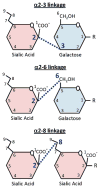
Sialic acid linkages. Sialic acids are added to the termini of glycans in an α2-3, α2-6, or α2-8 linkage. In the top two panels, sialic acid linkage to galactose is depicted, however other sugars, such as GalNAc, can be modified with sialic acid, depending upon the type of linkage. Note that the structures shown in the figure have been simplified (e.g., hydroxyl and acetyl groups are not represented)
Fig. 3
Fig. 3
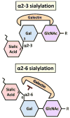
α2-6 sialylation blocks galectin binding. Galectins require a free hydroxyl group on the 6 carbon of galactose for binding [37], therefore α2-6 sialylation at this site inhibits galectin binding. In contrast, α2-3-linked sialic acids have variable effects on binding, depending upon the specific galectin species
Fig. 4
Fig. 4
Regulation of integrins by sialylation. α2-6 sialylation of N-linked glycans on the β1 integrin enhances cell adhesion to collagen I and stimulates migration and invasion
Fig. 5
Fig. 5
Sialyl Thomsen-nouvelle antigen. The sialyl Tn antigen is formed by α2-6 sialylation of GalNAc bound to serine or threonine
Fig. 6
Fig. 6
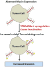
Sialyl Tn antigen expression correlates to increased invasion. Upregulation of the sialyltransferase, ST6GalNAc-I, or inactivation of the chaperone, Cosmc, contributes to an increase in sialyl Tn expression on O-linked glycans. Elevated levels of sialyl Tn antigen expressed on tumor cells is correlated to increased invasion; however, the mechanism remains unclear
Fig. 7
Fig. 7
Sialyl Lewis structures promote tumor dissemination. Sialyl Lewis structures on tumor cell glycoproteins interact with selectins expressed by activated endothelial cells, thereby facilitating tumor cell extravasation
Fig. 8
Fig. 8
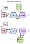
Sialyl Lewis antigens. Sialyl Lewis (sLe) antigens are tetrasaccharide structures composed of a GlcNAc-Gal backbone with fucose linked to GlcNAc and sialic acid α2-3 linked to Gal. sLex and sLea are different isomers, both of which bind to endothelial selectins
Fig. 9
Fig. 9
Inhibition of Fas-mediated apoptosis by α2-6 sialylation. α2-6 sialylation of the Fas receptor blocks apoptosis by preventing receptor internalization and formation of the death-inducing signaling complex (DISC)

References

    1. Lloyd KO, Old LJ. Human monoclonal antibodies to glycolipids and other carbohydrate antigens: dissection of the humoral immune response in cancer patients. Cancer Research. 1989;49:3445–3451. - PubMed
    1. Sell S. Cancer-associated carbohydrates identified by monoclonal antibodies. Human Pathology. 1990;21:1003–1019. - PubMed
    1. Varki A, Kannagi R, Toole BP. Glycosylation changes in cancer. In: Varki A, Cummings RD, Esko JD, Freeze HH, Stanley P, Bertozzi CR, Hart GW, Etzler ME, editors. Essentials of Glycobiology. 2. New York: Cold Spring Harbor Laboratory Press; 2009. pp. 580–670. - PubMed
    1. Hakomori S. Aberrant glycosylation in tumors and tumor-associated carbohydrate antigens. Advances in Cancer Research. 1989;52:257–331. - PubMed
    1. Zhou D, Levery SB, Hsu FF, Wang PG, Teneberg S, Almeida IC, Li Y, Xu H, Wang LX, Xia C, Ibrahim NK, Michael K. Immunologic mapping of glycomes: implications for cancer diagnosis and therapy. Frontiers in Bioscience (Scholar Edition) 2011;3:1520–1532. - PMC - PubMed

Publication types

LinkOut - more resources