. 2012 Jun 26;109(26):10504-9.
doi: 10.1073/pnas.1208314109. Epub 2012 Jun 13.

Hypoxia-inducible factor 1 transcriptional activity in endothelial cells is required for acute phase cardioprotection induced by ischemic preconditioning

Affiliations

Hypoxia-inducible factor 1 transcriptional activity in endothelial cells is required for acute phase cardioprotection induced by ischemic preconditioning

Kakali Sarkar et al. Proc Natl Acad Sci U S A. .

Abstract

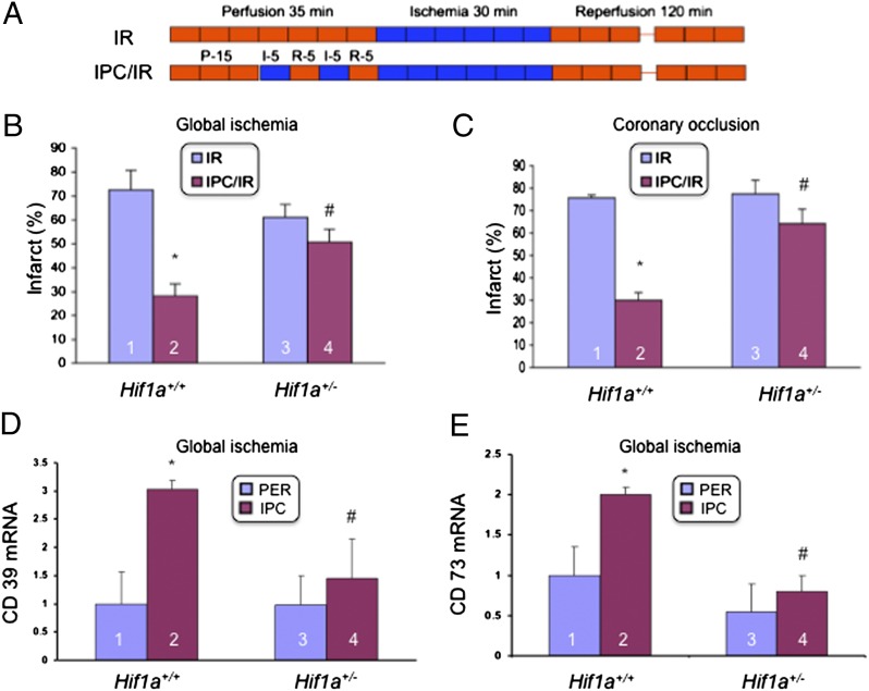
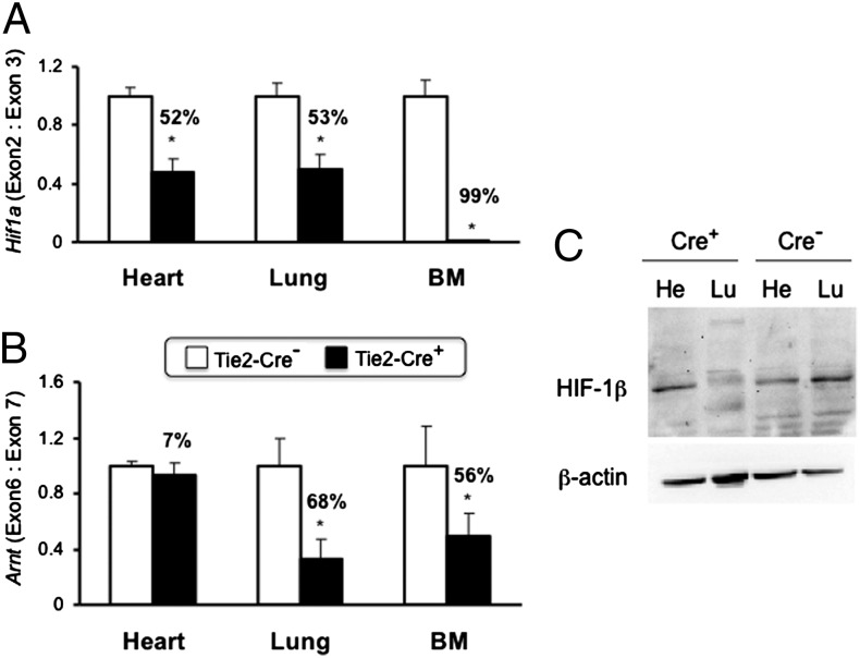
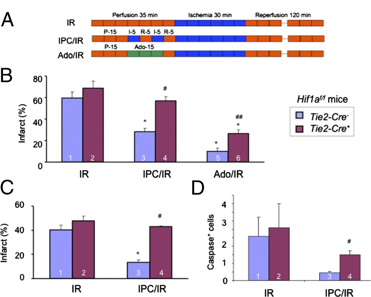
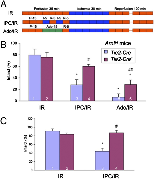
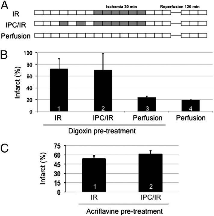
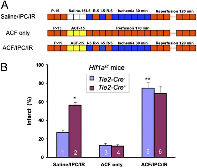
Infarction occurs when myocardial perfusion is interrupted for prolonged periods of time. Short episodes of ischemia and reperfusion protect against tissue injury when the heart is subjected to a subsequent prolonged ischemic episode, a phenomenon known as ischemic preconditioning (IPC). Hypoxia-inducible factor 1 (HIF-1) is a transcription factor that mediates adaptive responses to hypoxia/ischemia and is required for IPC. In this study, we performed a cellular and molecular characterization of the role of HIF-1 in IPC. We analyzed mice with knockout of HIF-1α or HIF-1β in Tie2(+) lineage cells, which include bone marrow (BM) and vascular endothelial cells, compared with control littermates. Hearts were subjected to 30 min of ischemia and 120 min of reperfusion, either as ex vivo Langendorff preparations or by in situ occlusion of the left anterior descending artery. The IPC stimulus consisted of two cycles of 5-min ischemia and 5-min reperfusion. Mice lacking HIF-1α or HIF-1β in Tie2(+) lineage cells showed complete absence of protection induced by IPC, whereas significant protection was induced by adenosine infusion. Treatment of mice with a HIF-1 inhibitor (digoxin or acriflavine) 4 h before Langendorff perfusion resulted in loss of IPC, as did administration of acriflavine directly into the perfusate immediately before IPC. We conclude that HIF-1 activity in endothelial cells is required for acute IPC. Expression and dimerization of the HIF-1α and HIF-1β subunits is required, suggesting that the heterodimer is functioning as a transcriptional activator, despite the acute nature of the response.


Conflict of interest statement

The authors declare no conflict of interest.

Figures

Fig. 1.
Fig. 1.
Loss of the cardioprotective effect of ischemic preconditioning in Hif1a+/− mice. (A) Hearts were subjected to 30 min of ischemia and 120 min of reperfusion, either alone (IR) or preceded by ischemic preconditioning (IPC), consisting of two cycles of 5 min of ischemia and 5 min of reperfusion (IPC/IR). (B and C) Effect of IPC on infarct size (infarct area/total area at risk × 100%; mean ± SD, n = 3 hearts in each group), as determined by staining with 1% triphenyltetrazolium chloride (TTC), was analyzed in ex vivo Langendorff-perfused hearts (B) or in hearts subjected to in situ coronary artery occlusion (C). *P < 0.01 vs. column 1; #P < 0.01 vs. column 2 (Student’s t test). (D and E) Hearts isolated from Hif1a+/+ and Hif1a+/− mice were either subjected to IPC or perfused continuously (PER) for 40 min and total RNA was isolated and analyzed for the expression of CD39 (D) and CD73 (E) mRNA by quantitative real-time RT-PCR (mean ± SEM; n = 5 hearts in each group). *P < 0.05 vs. column 1; #P < 0.05 vs. column 2 (Student’s t test).
Fig. 2.
Fig. 2.
Analysis of gene deletion in conditional knockout mice. (A and B) DNA was isolated from heart, lung, and bone marrow of Hif1af/f (A) and Arntf/f (B) mice that were either Tie2-Cre (open bars) or Tie2-Cre+ (closed bars). Efficiency of gene deletion was determined by quantitative real-time PCR as the ratio of the targeted exon to the next downstream exon × 100%. *P < 0.05 vs. Tie2-Cre (n = 4–6; Student’s t test on log-converted ratios). (C) Immunoblot assay of HIF-1β and β-actin protein levels in heart (He) and lung (Lu) tissue lysates from Arntf/f; Tie2-Cre+ and Arntf/f; Tie2-Cre mice.
Fig. 3.
Fig. 3.
Effect of IPC or adenosine perfusion in hearts of Hif1af/f;Tie2-Cre mice. (A–C) Hearts were subjected to 30 min of ischemia and 120 min of reperfusion (IR), ischemic preconditioning consisting of two cycles of 5-min ischemia + 5-min reperfusion followed by IR (IPC/IR), or 200-μM adenosine infusion for 15 min followed by IR (Ado/IR) in either the ex vivo Langendorff perfusion (B) or in situ coronary artery occlusion (C) model. Infarct size (mean ± SEM) was determined by TTC staining. *P < 0.05 vs. column 1; #P < 0.05 vs. column 3; ##P < 0.05 vs. column 5 (n = 3–4, Student’s t test). (D) Hearts were subjected to IR and IPC/IR as described except that reperfusion was for 45 min rather than 120 min and heart sections were analyzed by immunohistochemistry using an antibody specific for cleaved (activated) caspase 3. #P < 0.05 vs. column 3 (n = 3; Student’s t test).
Fig. 4.
Fig. 4.
Effect of IPC or adenosine perfusion in hearts of Arntf/f; Tie2-Cre mice. (A–C) Hearts were subjected to 30 min of ischemia and 120 min of reperfusion (IR), ischemic preconditioning consisting of two cycles of 5-min ischemia + 5-min reperfusion followed by IR (IPC/IR), or 200-μM adenosine infusion for 15 min followed by IR (Ado/IR) in either the ex vivo Langendorff perfusion (B) or in situ coronary artery occlusion (C) model and infarct size (mean ± SEM; n = 3 hearts in each group) was determined by TTC staining. *P < 0.05 vs. column 1; #P < 0.05 vs. column 3; ##P < 0.05 vs. column 5 (Student’s t test).
Fig. 5.
Fig. 5.
Effect of parenteral administration of HIF-1 inhibitors on cardioprotection mediated by IPC. (AC) Wild-type mice were pretreated with an i.p. injection of digoxin (B) or acriflavine (C) at a dose of 2 mg/kg. Four hours later, Langendorff heart preparations were subjected to IR, IPC/IR, or perfusion (without IPC or IR) and infarct size was determined by TTC staining (mean ± SEM, n = 3–4 hearts in each group).
Fig. 6.
Fig. 6.
Effect of acriflavine infusion immediately before IPC/IR on cardioprotection. (A and B) Langendorff heart preparations from Hif1af/f; Tie2-Cre and Hif1af/f; Tie2-Cre+ mice were perfused with saline or 1 μM acriflavine (ACF) for 15 min before IPC/IR or perfusion only (A) and infarct size (B) was determined by TTC staining (mean ± SEM, n = 3). *P < 0.05, **P < 0.01 vs. column 1; Student’s t test.

Similar articles

Cited by

References

    1. Lopaschuk GD, Kelly DP. Signalling in cardiac metabolism. Cardiovasc Res. 2008;79:205–207. - PubMed
    1. Nallamothu BK, Bradley EH, Krumholz HM. Time to treatment in primary percutaneous coronary intervention. N Engl J Med. 2007;357:1631–1638. - PubMed
    1. Eltzschig HK, Eckle T. Ischemia and reperfusion—from mechanism to translation. Nat Med. 2011;17:1391–1401. - PMC - PubMed
    1. Murry CE, Jennings RB, Reimer KA. Preconditioning with ischemia: A delay of lethal cell injury in ischemic myocardium. Circulation. 1986;74:1124–1136. - PubMed
    1. Kuzuya T, et al. Delayed effects of sublethal ischemia on the acquisition of tolerance to ischemia. Circ Res. 1993;72:1293–1299. - PubMed

Publication types

LinkOut - more resources