. 2012 Jul 3;109(27):E1868-77.
doi: 10.1073/pnas.1116889109. Epub 2012 Jun 13.

Notch ligand delta-like 4 blockade attenuates atherosclerosis and metabolic disorders

Affiliations

Notch ligand delta-like 4 blockade attenuates atherosclerosis and metabolic disorders

Daiju Fukuda et al. Proc Natl Acad Sci U S A. .

Abstract

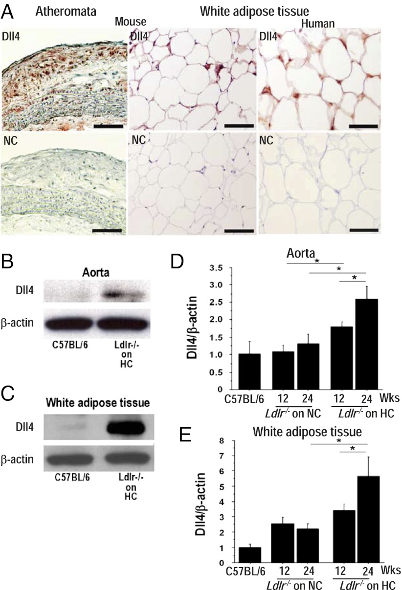
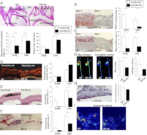
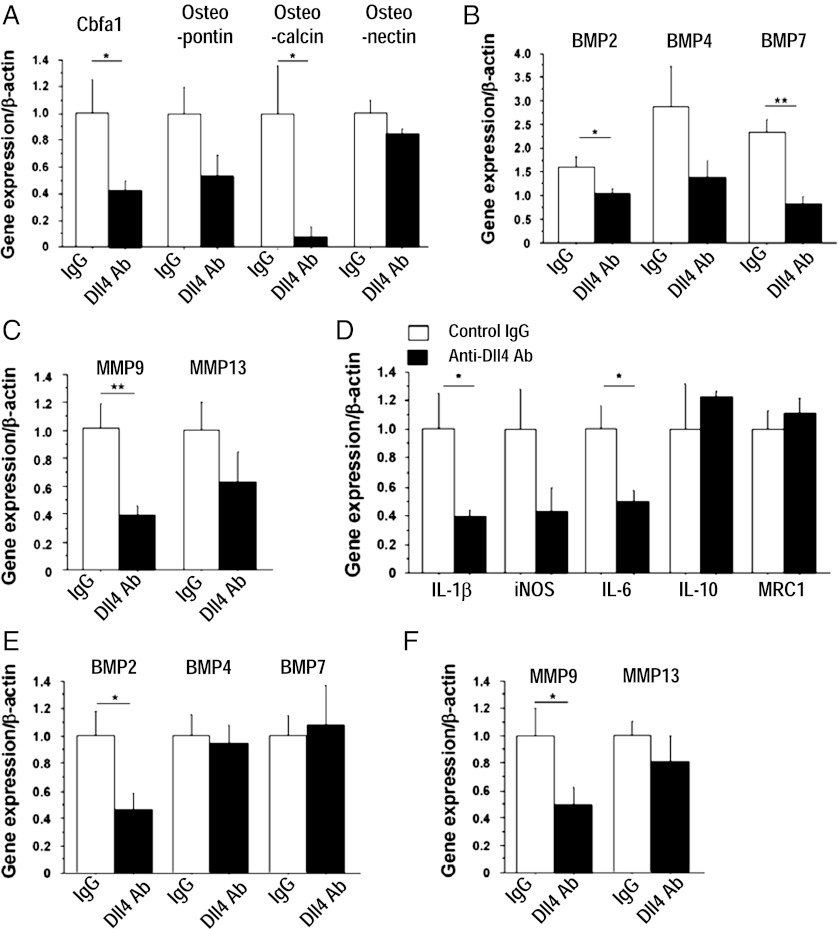
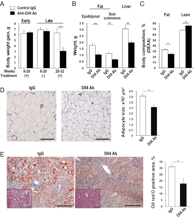
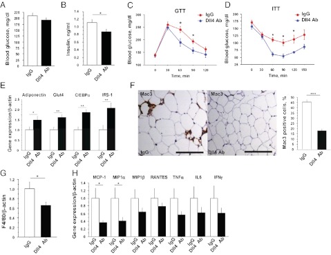
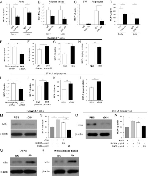
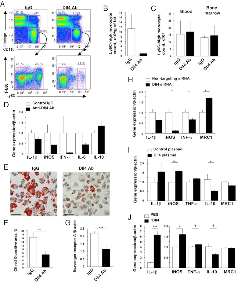
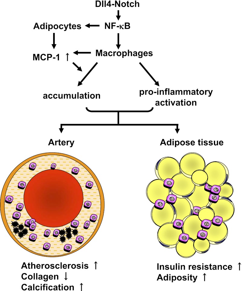
Atherosclerosis and insulin resistance are major components of the cardiometabolic syndrome, a global health threat associated with a systemic inflammatory state. Notch signaling regulates tissue development and participates in innate and adaptive immunity in adults. The role of Notch signaling in cardiometabolic inflammation, however, remains obscure. We noted that a high-fat, high-cholesterol diet increased expression of the Notch ligand Delta-like 4 (Dll4) in atheromata and fat tissue in LDL-receptor-deficient mice. Blockade of Dll4-Notch signaling using neutralizing anti-Dll4 antibody attenuated the development of atherosclerosis, diminished plaque calcification, improved insulin resistance, and decreased fat accumulation. These changes were accompanied by decreased macrophage accumulation, diminished expression of monocyte chemoattractant protein-1 (MCP-1), and lower levels of nuclear factor-κB (NF-κB) activation. In vitro cell culture experiments revealed that Dll4-mediated Notch signaling increases MCP-1 expression via NF-κB, providing a possible mechanism for in vivo effects. Furthermore, Dll4 skewed macrophages toward a proinflammatory phenotype ("M1"). These results suggest that Dll4-Notch signaling plays a central role in the shared mechanism for the pathogenesis of cardiometabolic disorders.


Conflict of interest statement

The authors declare no conflict of interest.

Figures

Fig. 1.
Fig. 1.
Characterization of Dll4 expression and effects of Dll4 blockade. (A) Immunostaining of Dll4 in atheroma (Upper Left) and epididymal fat (Upper Center) obtained from 32-wk-old Ldlr−/− mice on a high-fat and high-cholesterol (HC) diet and in human white adipose tissue (Upper Right). (Lower) Staining with nonimmune IgG (NC). (Scale bar: 100 μm.) (B and C) Immunoblotting of Dll4 in aortas (B) and white adipose tissue (C). (D and E) Effect of HC diet on Dll4 RNA expression in aortas (D) and adipose tissue (E) of Ldlr−/− mice. Mice were fed a HC diet from 8 wk of age for 12 or 24 wk. HC diet promotes Dll4 expression. Longer consumption of HC diet enhances the effect. NC, normal chow. A and B, n = 4. D and E, n = 4. *P < 0.05; **P < 0.01. All values are mean ± SEM.
Fig. 2.
Fig. 2.
Effects of Dll4 blockade on atherogenesis. (A) H&E staining of the aortic arch. (Scale bar: 200 μm.) (B–F) Immunostaining for MCP-1 (B) and Mac3 (C), picrosirius red staining (D), von Kossa staining (E), and alkaline phosphatase (ALP) activity (F) in plaques. (Scale bars: 100 μm.) (G) Simultaneous ex vivo mapping using fluorescence reflectance imaging of aortas (late-phase treatment, n = 4). (H) ALP activity and thickness of aortic valves. (Scale bar: 10 μm.) (I) Ex vivo fluorescence reflectance imaging of aortic valves. A–C and E show the early-phase treatment (n = 7–8). D and F show the late-phase treatment (n = 14–15). *P < 0.05; **P < 0.01. All values are mean ± SEM.
Fig. 3.
Fig. 3.
Role of Dll4 in the expression of osteogenic regulators, MMPs, and inflammatory molecules. (A–C) Quantitative RT-PCR analysis of osteogenic regulators (A), BMPs (B), and MMPs (C) in aortas. (D) Quantitative RT-PCR analyses of the expression of inflammatory molecules in aortas. (E and F) Quantitative RT-PCR analyses of BMPs (E) and MMPs (F) in peritoneal macrophages. A–F show early-phase treatment (n = 7–8). *P < 0.05; **P < 0.01. All values are mean ± SEM.
Fig. 4.
Fig. 4.
Effects of Dll4 blockade on fat accumulation. (A) Differences in body weight gain during the study periods (early-phase treatment, n = 7–8; late-phase treatment, n = 19–20). (B) Weight of fat and liver (late-phase treatment, n = 19–20). (C) Results of dual-energy X-ray absorptiometry (DEXA), n = 5–6. (D) H&E staining and quantification of adipocyte size in epididymal fat (n = 7). (Scale bar: 200 μm.) (E) Oil red O staining of the liver and quantification of lipid deposition (late-phase treatment, n = 9–10). (Insets) H&E staining. (Scale bar: 100 μm.) *P < 0.05; **P < 0.01; ***P < 0.001. All values are mean ± SEM.
Fig. 5.
Fig. 5.
Effects of Dll4 blockade on insulin sensitivity and macrophage accumulation in fat tissue. (A and B) Blood glucose levels (A) and serum insulin levels (B) after 4-h fasting (n = 9). (C and D) Glucose tolerance test (GTT) after 16-h fasting and insulin tolerance test (ITT) after 4-h fasting (n = 7). (E) Quantitative RT-PCR analyses of genes related to insulin sensitivity in epididymal fat (n = 9–10). (F) Mac3 staining and population of Mac3-positive cells in epididymal fat (n = 7). (Scale bar: 100 μm.) (G) Quantitative RT-PCR analysis of F4/80 in epididymal fat (n = 9–10). (H) Quantitative RT-PCR analyses of expression of chemokines and cytokines in epididymal fat (n = 9–10). All data are from late-phase treatment. *P < 0.05; **P < 0.01; ***P < 0.001. All values are mean ± SEM.
Fig. 6.
Fig. 6.
Role of Dll4 in MCP-1 expression in vivo and in vitro. (A and B) MCP-1 RNA expression in aortas (A) and fat tissue (B). (C) MCP-1 RNA expression in SVF and adipocytes obtained from epididymal fat. (D) Serum MCP-1 levels. Early-phase treatment, n = 7–8; late-phase treatment, n = 9–10. (E–G) Effects of RNAi silencing of Dll4 (E), transfection of plasmid encoding Dll4 (F), and stimulation with immobilized rDll4 (G) on MCP-1 RNA expression in RAW264.7 cells. (H) MCP-1 protein levels in supernatant of rDll4-treated RAW264.7 cells. (I–K) Effects of RNAi silencing of Dll4 (I), transfection of plasmid encoding Dll4 (J), and stimulation with immobilized rDll4 (K) on MCP-1 RNA expression in 3T3-L1 adipocytes. (L) MCP-1 protein levels in supernatant of rDll4-treated 3T3-L1 adipocytes. (M) Degradation of IκBα in rDll4-stimulated RAW264.7 cells. (N) Reduction of MCP-1 expression by NF-κB inhibitor SN50 in rDll4-stimulated RAW264.7 cells. SN50M, inactive control for SN50. (O) Degradation of IκBα in rDll4-stimulated 3T3-L1 adipocytes. (P) Reduction of MCP-1 expression by NF-κB inhibitor SN50 in rDll4-stimulated 3T3-L1 adipocytes. (Q and R) Inhibition of NF-κB in aortas (Q) and fat tissues (R) determined by degradation of IκBα in Dll4 Ab-treated Ldlr−/− mice. E–P, n = 6. Q and R show representative data from late-phase treatment, n = 4. *P < 0.05; **P < 0.01. All values are mean ± SEM.
Fig. 7.
Fig. 7.
Role of Dll4 in proinflammatory activation of macrophages. (A) Flow cytometry analyses of SVF obtained from epididymal fat. (B) Count of Ly6C-high monocytes in fat. (C) Ly6C-high monocyte population in blood and bone marrow. (D) Quantitative RT-PCR analyses of expression of inflammatory molecules in F4/80-positive macrophages obtained from fat. (E and F) Oil red O staining (E) and quantification of lipid deposition (F) of peritoneal macrophages obtained from Ldlr−/− mice that received late-phase treatment. (G) Quantitative RT-PCR analyses of expression of macrophage scavenger receptor-A in peritoneal macrophages. (H–J) Effects of RNAi silencing of Dll4 (H), overexpression of Dll4 using expressing plasmid (I), and stimulation with immobilized rDll4 (J) on the expression of inflammatory molecules in RAW264.7 cells. A–C, n = 3; D–G, n = 9–10; H–J, n = 6. P = 0.05; *P < 0.05; **P < 0.01; ***P < 0.001. All values are mean ± SEM.
Fig. P1.
Fig. P1.
Role of the Dll4-Notch axis in cardiometabolic disorders. The Dll4-Notch axis activates the NF-κB pathway. Activation of NF-κB increases the expression of MCP-1, which leads to the accumulation and proinflammatory activation of macrophages in organs such as arteries and adipose tissue. Accumulation of activated macrophages contributes to the pathogenesis of cardiometabolic disorders, including atherosclerotic vascular diseases and insulin resistance.

References

    1. Eckel RH, Grundy SM, Zimmet PZ. The metabolic syndrome. Lancet. 2005;365:1415–1428. - PubMed
    1. Hotamisligil GS. Inflammation and metabolic disorders. Nature. 2006;444:860–867. - PubMed
    1. Artavanis-Tsakonas S, Rand MD, Lake RJ. Notch signaling: Cell fate control and signal integration in development. Science. 1999;284:770–776. - PubMed
    1. Aster JC, Pear WS, Blacklow SC. Notch signaling in leukemia. Annu Rev Pathol. 2008;3:587–613. - PMC - PubMed
    1. Radtke F, Fasnacht N, Macdonald HR. Notch signaling in the immune system. Immunity. 2010;32:14–27. - PubMed

Publication types

MeSH terms