. 2012 Jun 13;32(24):8127-37.
doi: 10.1523/JNEUROSCI.6034-11.2012.

Brain-derived neurotrophic factor activation of CaM-kinase kinase via transient receptor potential canonical channels induces the translation and synaptic incorporation of GluA1-containing calcium-permeable AMPA receptors

Affiliations

Brain-derived neurotrophic factor activation of CaM-kinase kinase via transient receptor potential canonical channels induces the translation and synaptic incorporation of GluA1-containing calcium-permeable AMPA receptors

Dale A Fortin et al. J Neurosci. .

Abstract

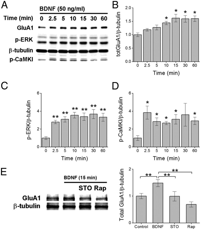
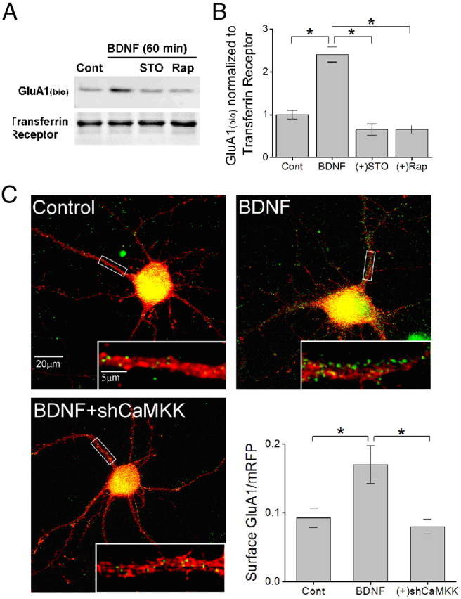
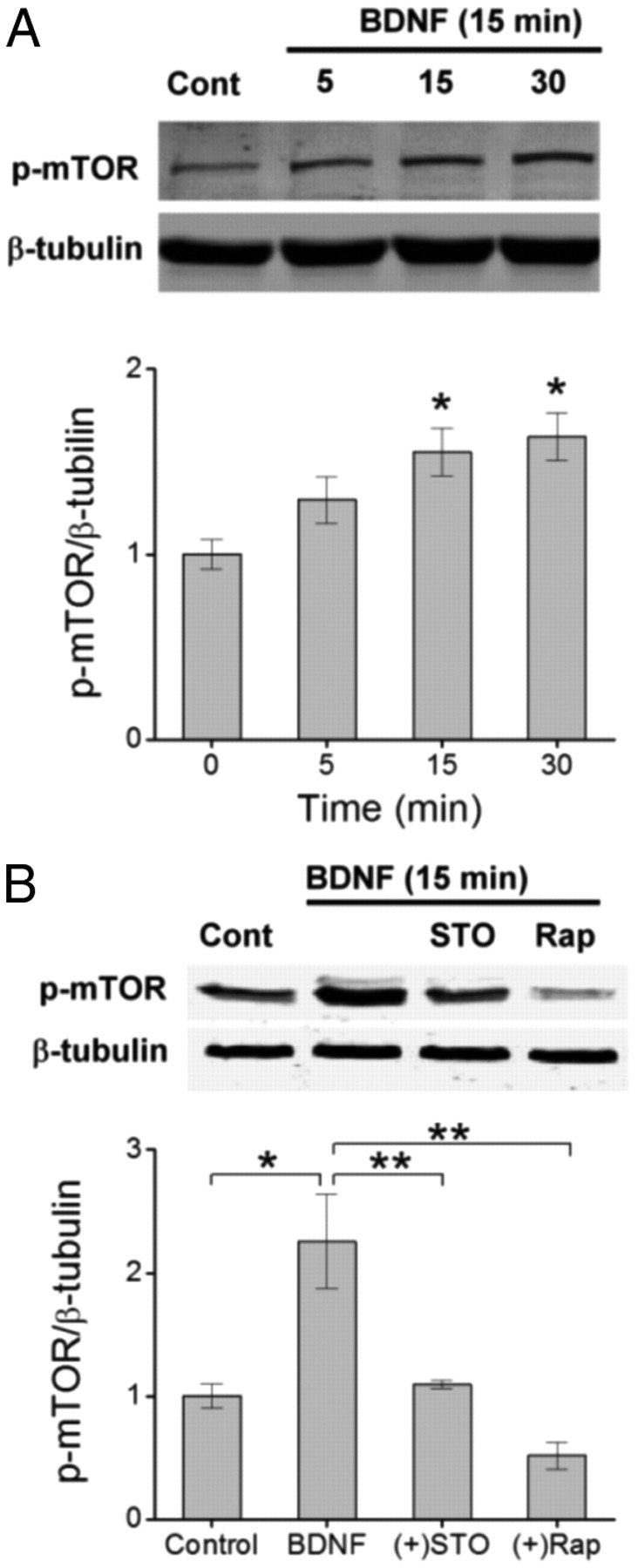
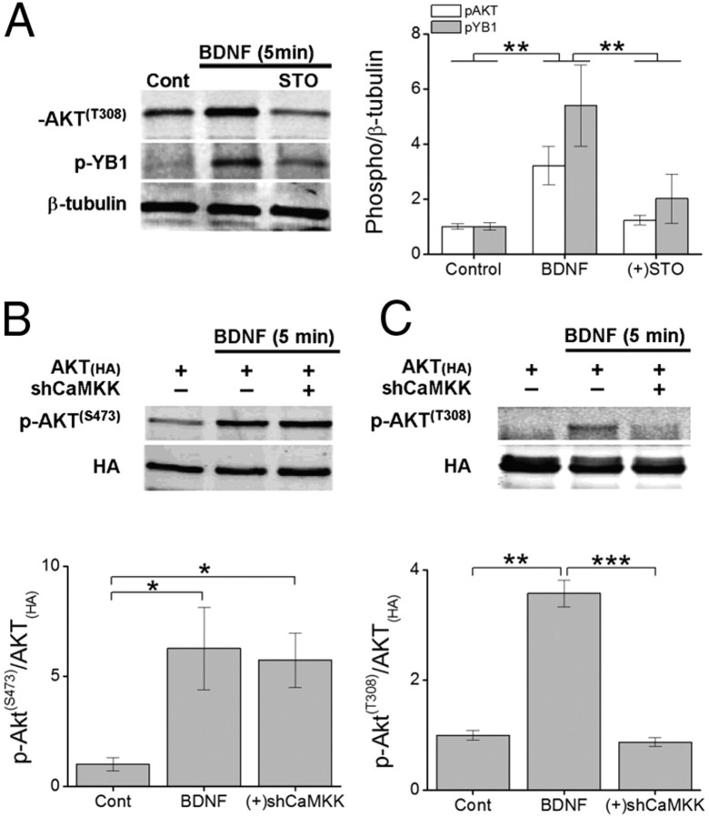
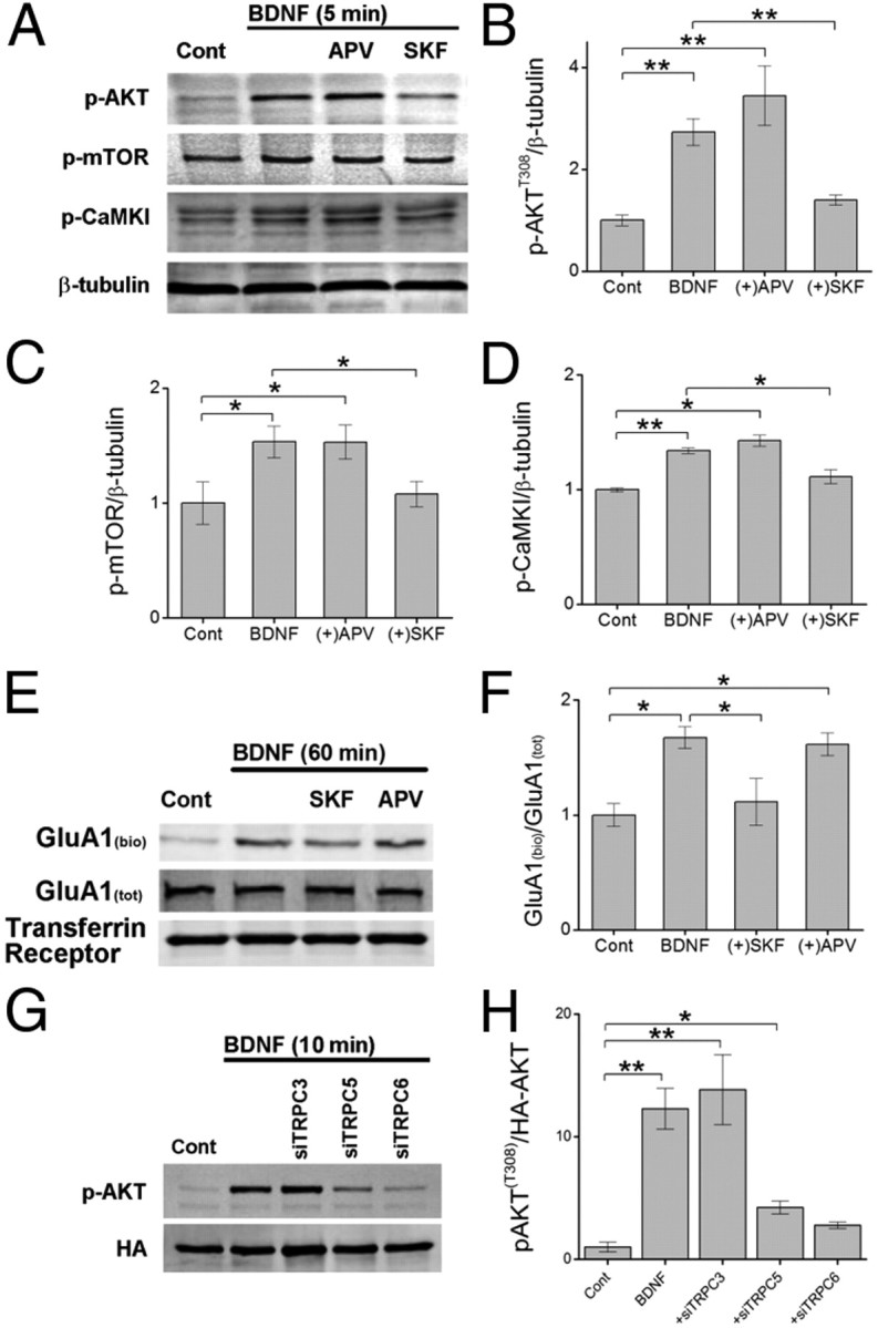
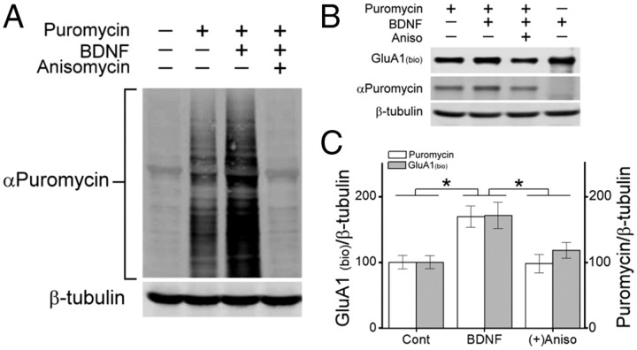
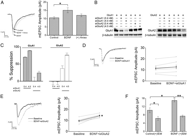
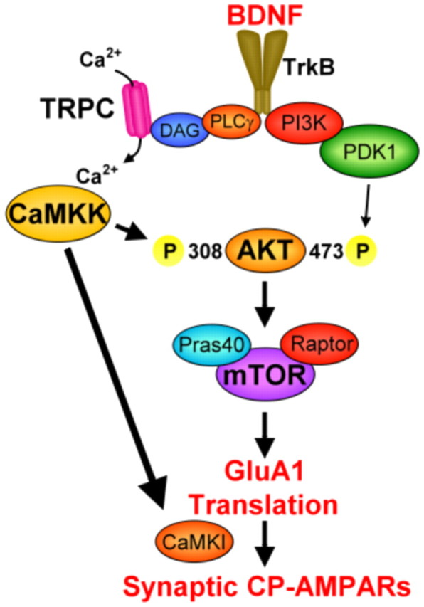
Glutamatergic synapses in early postnatal development transiently express calcium-permeable AMPA receptors (CP-AMPARs). Although these GluA2-lacking receptors are essential and are elevated in response to brain-derived neurotrophic factor (BDNF), little is known regarding molecular mechanisms that govern their expression and synaptic insertion. Here we show that BDNF-induced GluA1 translation in rat primary hippocampal neurons requires the activation of mammalian target of rapamycin (mTOR) via calcium calmodulin-dependent protein kinase kinase (CaMKK). Specifically, BDNF-mediated phosphorylation of threonine 308 (T308) in AKT, a known substrate of CaMKK and an upstream activator of mTOR-dependent translation, was prevented by (1) pharmacological inhibition of CaMKK with STO-609, (2) overexpression of a dominant-negative CaMKK, or (3) short hairpin-mediated knockdown of CaMKK. GluA1 surface expression induced by BDNF, as assessed by immunocytochemistry using an extracellular N-terminal GluA1 antibody or by surface biotinylation, was impaired following knockdown of CaMKK or treatment with STO-609. Activation of CaMKK by BDNF requires transient receptor potential canonical (TRPC) channels as SKF-96365, but not the NMDA receptor antagonist d-APV, prevented BDNF-induced GluA1 surface expression as well as phosphorylation of CaMKI, AKT(T308), and mTOR. Using siRNA we confirmed the involvement of TRPC5 and TRPC6 subunits in BDNF-induced AKT(T308) phosphorylation. The BDNF-induced increase in mEPSC was blocked by IEM-1460, a selected antagonist of CP-AMPARs, as well as by the specific repression of acute GluA1 translation via siRNA to GluA1 but not GluA2. Together these data support the conclusion that newly synthesized GluA1 subunits, induced by BDNF, are readily incorporated into synapses where they enhance the expression of CP-AMPARs and synaptic strength.


Figures

Figure 1.
Figure 1.
BDNF-induced increase in total GluA1 protein requires CaMKK. A–D, Representative immunoblot of hippocampal cell lysates illustrating the increase in total GluA1 (A, B), phosphorylated ERK (p-ERK) (C), and CaMKK-mediated phosphorylation of CaMKI (p-CaMKI) (D) following increasing times of BDNF treatment. Error bars indicate SEM (n = 5 from 5 independent experiments). *p < 0.05 by one-way ANOVA and Tukey's post hoc test. E, Representative immunoblot (left) and quantification (4 independent experiments) of total GluA1 protein following 15 min of BDNF treatment in the absence or presence of 10 μm STO-609 (STO, CaMKK inhibitor) or 1 μm rapamycin (Rap, mTORC1 inhibitor). Error bars indicate SEM. *p < 0.05, **p < 0.01 by Student's t test. In A and E, β-tubulin is shown as loading control.
Figure 2.
Figure 2.
CaMKK mediates BDNF-induced increases in GluA1 surface expression. A, Representative immunoblot of biotinylated surface GluA1 (GluA1bio) and surface transferrin receptor following 60 min of BDNF treatment in the absence or presence of STO-609 or Rap as shown. B, Quantification of surface biotinylated GluA1 normalized to surface transferrin receptor for indicated conditions (n = 5 from 5 independent experiments). C, Representative immunofluorescence images of hippocampal primary neurons (10 DIV) transfected with monomeric RFP (red) and superimposed with surface GluA1 (pseudo-colored in green) for control. Neurons treated with 50 ng/ml BDNF plus or minus coexpression of short hairpins to α and β CaMKK (shCaMKK). Inset shows a magnification of the boxed segment of proximal dendrite. Lower right, Group data for surface GluA1 levels for indicated conditions (n = 8–12 neurons per coverslip from 3 independent experiments). Error bars indicate SEM *p < 0.05 by Student's t test.
Figure 3.
Figure 3.
CaMKK functions upstream to activate mTOR. A, Top, Representative Western blot illustrating the time-dependent increase in phosphorylated mTOR (p-mTOR, S2448) following treatment with BDNF for the indicated times shown. Bottom, Group data for the ratio of p-mTOR to β-tubulin for each given time point. Error bars indicate SEM (n = 3 from 3 independent experiments). *p < 0.05 by one-way ANOVA and Tukey's post hoc test. B, Top, Immunoblot of p-mTOR levels following BDNF treatment for 15 min in the absence or presence of STO-609 (10 μm) or rapamycin (1 μm). Bottom, Group data for p-mTOR levels normalized to β-tubulin for indicated conditions. Error bars indicate SEM (n = 4 from 4 independent experiments). **p < 0.01 by Student's t test.
Figure 4.
Figure 4.
BDNF-induced phosphorylation of AKT at T308, but not S473, requires CaMKK. A, Left, Representative Western blot illustrating the change in phosphorylation of AKT [p-AKT(T308)] and its substrate Y-box binding protein 1 (p-YB1) following 5 min of BDNF treatment in presence or absence of STO-609. Right, Group data for both p-AKT and p-YB1 normalized to β-tubulin (n = 4 from 4 independent experiments). B, C, Representative Western blots of immunoprecipitated HA-tagged AKT expressed or coexpressed with shCaMKK in hippocampal neurons treated with BDNF for 5 min and probed for phosphorylation at AKTT308 (B) or AKTS473 (C). Group data of p-AKT for T308 or S473 (n = 3 from 3 independent experiments) to total AKT for conditions indicated are shown below. Error bars indicate SEM. *p < 0.05, **p < 0.01, ***p < 0.001 by Student's t test.
Figure 5.
Figure 5.
TRPC channels contribute to BDNF-mediated activation of CaMKK, the mTOR complex, and membrane trafficking of GluA1. A–D, Representative Western blot of hippocampal cell lysates illustrating the increase in phosphorylated AKT (T308), mTOR (S2448), and CaMKI (T177) following 5 min of BDNF treatment in the absence or presence of APV (25 μm or SKF-96265 (SKF, 30 μm) (A). Quantification of changes in total p-AKT (B), p-mTOR (C), and p-CaMKI (D) normalized to β-tubulin for indicated conditions. Error bars indicate SEM (n = 5 from 5 independent experiments). E, Representative Western blot of surface biotinylated GluA1 [GluA1(bio)], GluA1(tot), and transferrin receptor following 60 min of BDNF treatment in the presence or absence of SKF or APV. F, Mean data from four independent experiments for conditions shown. G, Neurons were transfected with HA-AKT alone or with siTRPC3, siTRPC5, or siTRPC6. After 72 h, neurons were treated with BDNF for 10 min before undergoing immunoprecipitation to obtain HA-AKT. Representative Western blot of pAKT (T308) and HA following BDNF treatment is shown for indicated conditions. H, Group data for conditions shown in E demonstrating loss of AKTT308 phosphorylation by BDNF in the presence of siTRPC5 or siTRPC6. *p < 0.05, **p < 0.01 by Student's t test.
Figure 6.
Figure 6.
BDNF induces surface delivery of newly translated GluA1. A, Immunoblot of hippocampal total cell lysates following a 10 min puromycin (10 ng/ml) pulse followed by BDNF stimulation (40 min) in the absence or presence of anisomycin (Aniso, 10 μm) and probed with puromycin or β-tubulin antibodies. B, Western blot showing levels of surface biotinylated GluA1 (streptavidin), and puromycin incorporation (puromycin) from hippocampal lysates immunoprecipitated with an antibody to GluA1 following BDNF stimulation in the absence or presence of anisomycin. β-Tubulin was probed from accompanying total cell lysates. C, Quantification of the ratio of surface biotinylated GluA1 and puromycin incorporation to β-tubulin for indicated conditions (n = 5 from 5 independent experiments). Error bars indicate SEM. *p < 0.05 by Student's t test.
Figure 7.
Figure 7.
BDNF-induced increase in synaptic strength requires protein synthesis of GluA1 subunits coupled to expression of CP-AMPARs. A, Left, Example traces of mEPSCs recorded at room temperature at a holding potential of −70 mV for each condition indicated. Traces were an average of 100–150 consecutive events recorded during each treatment condition. In the indicated experiments, neurons were pretreated with anisomycin for 30 min before the addition of BDNF to impair translation. Right, Group data for mean mEPSC amplitude for control (n = 12), BDNF (n = 6; *p < 0.05 by Student's t test) and BDNF in the presence of anisomycin (Aniso; n = 6). B, Representative Western blot for GluA1 and GluA2 subunits expressed in HEK cells in the absence or presence of 0.4 or 4.0 nm siRNA for GluA1 or GluA2. β-Tubulin was used as a loading control. C, Quantification of GluA1 and GluA2 knockdown for conditions indicated in B for four independent experiments. D, E, Left, Average mEPSCs recorded during baseline and following treatment with BDNF with inclusion of siGluA1 (D) or siGluA2 (E) in the patch pipette. Right, Individual (gray) and mean (black) mEPSC amplitudes for siGluA1 (D; n = 7) and siGluA2 (E; n = 5). *p < 0.05 by Student's paired t test. F, Group data illustrating the mean reduction in mEPSC amplitude as a percentage of baseline responses following IEM treatment under baseline conditions (left; n = 12) and following BDNF stimulation (right; n = 7). Error bars indicate SEM. *p < 0.05 by Student's paired t test.
Figure 8.
Figure 8.
Schematic of signaling pathway involving CaMKK regulation of GluA1 synthesis and synaptic incorporation as CP-AMPARs. Signaling shown in bold font is documented in the current report whereas signaling in normal font is taken from the literature as cited in the text.

References

    1. Aizenman CD, Muñoz-Elías G, Cline HT. Visually driven modulation of glutamatergic synaptic transmission is mediated by the regulation of intracellular polyamines. Neuron. 2002;34:623–634. - PubMed
    1. Alessi DR, Andjelkovic M, Caudwell B, Cron P, Morrice N, Cohen P, Hemmings BA. Mechanism of activation of protein kinase B by insulin and IGF-1. EMBO J. 1996;15:6541–6551. - PMC - PubMed
    1. Alessi DR, James SR, Downes CP, Holmes AB, Gaffney PR, Reese CB, Cohen P. Characterization of a 3-phosphoinositide-dependent protein kinase which phosphorylates and activates protein kinase Balpha. Curr Biol. 1997;7:261–269. - PubMed
    1. Alsina B, Vu T, Cohen-Cory S. Visualizing synapse formation in arborizing optic axons in vivo: dynamics and modulation by BDNF. Nat Neurosci. 2001;4:1093–1101. - PubMed
    1. Amaral MD, Pozzo-Miller L. TRPC3 channels are necessary for brain-derived neurotrophic factor to activate a nonselective cationic current and to induce dendritic spine formation. J Neurosci. 2007a;27:5179–5189. - PMC - PubMed

Publication types

MeSH terms

Substances