. 2013 Jan 1;521(1):203-12.
doi: 10.1002/cne.23171.

Neurotropic virus tracing suggests a membranous-coating-mediated mechanism for transsynaptic communication

Affiliations

Neurotropic virus tracing suggests a membranous-coating-mediated mechanism for transsynaptic communication

Yan-Chao Li et al. J Comp Neurol. .

Abstract

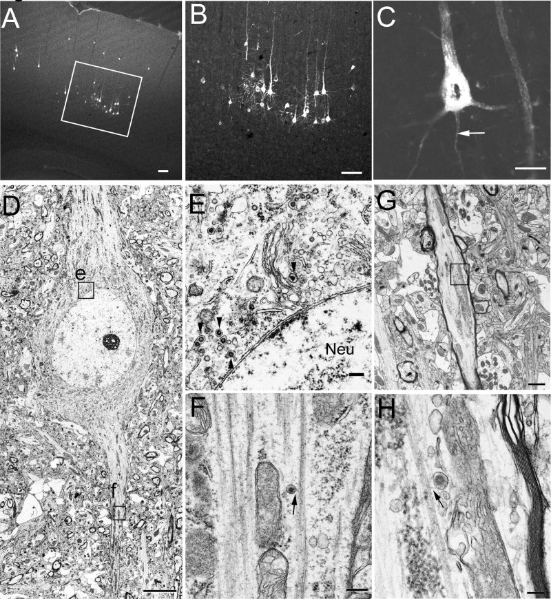
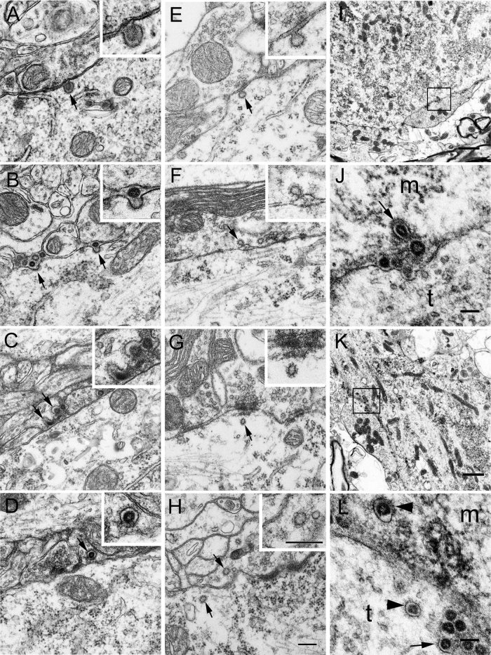
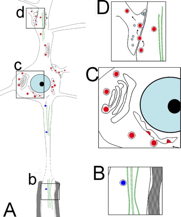
Swine hemagglutinating encephalomyelitis virus (HEV) has been shown to have a capability to propagate via neural circuits to the central nervous system after peripheral inoculation, resulting in acute deadly encephalomyelitis in natural host piglets as well as in experimental younger rodents. This study has systematically examined the assembly and dissemination of HEV 67N in the primary motor cortex of infected rats and provides additional evidence indicating that membranous-coating-mediated endo-/exocytosis can be used by HEV for its transsynaptic transfer. In addition, our results suggested that this transsynaptic pathway could adapted for larger granular materials, such as viruses. These findings should help in understanding the mechanisms underlying coronavirus infections as well as the intercellular exchanges occurring at the synaptic junctions.


Figures

Figure 1
Figure 1
HEV‐infected neurons in the primary motor cortex at day 4 p.i. A–C: Fluorescence micrographs showing HEV‐infected neurons in the primary motor cortex at day 4 p.i. A is a low‐magnification micrograph, and the boxed area is enlarged in B, showing that infected neurons are located mainly in layer V of the primary motor cortex. C is an image of a 0.6‐μm‐thick optical section, showing the punctuate cytoplasmic staining of HEV, which is distributed throughout the cell body, dendrites, and axon (arrow). D–H: EM photomicrographs showing HEV particles in infected pyramidal cells. D shows an infected pyramidal cell, in which both progeny virions and budding profiles are observed in the somatodendritic cytoplasm. The boxed area e is enlarged in E, showing virus particles present in small vesicles (arrowheads) in the perikaryon of the pyramidal cell. No virus particles are found in the nuclei (Neu). The boxed area f is enlarged in F, showing an HEV particle within a small vesicle (arrow), which is closely associated with the fasciculated microtubules in the initial segment. G shows a longitudinally cut myelinated axon of an infected pyramidal cell. The boxed area is enlarged in H, showing a virus‐containing vesicle (arrow) closely associated with the microtubules. Scale bars = 100 μm in A,B; 20 μm in C; 5 μm in D; 200 nm in E,F; 1 μm in G; 100 nm in H.
Figure 2
Figure 2
EM photomicrographs showing the replication and assembly of HEV in pyramidal cells. Viral assembly inside the infected cells is initiated by an electron‐dense crescent segment (arrows in A), which is attached to the external membrane of the ER cistern. Larger crescent segments (arrows in A,B) bulge into the lumen of the ER cistern by incorporation of the lipid bilayer of the ER into the viral envelope. Short stalks (arrows in B) are found still continuing with the ER membrane. The virus particles in the lumen of the ER cistern are covered with well‐defined surface projections (C). D shows virus particles in Golgi and ER areas in the perikaryal cytoplasm of a pyramidal cell. No membranous decorations are present on the outer surface of the virus‐containing vesicle near the ER cistern (indicated by f, and enlarged in F). By contrast, the virion in the Golgi area (indicated by g, and enlarged in G) is enclosed within a vesicle with spinule coats on the outer surface. In both cases, a layer of surface projections is seen surrounding the viral envelope beneath the vesicle membrane (F,G). E shows a Golgi apparatus in the primary dendrite of a proximal cell, where viral budding profiles are observed in the lateral rims of the Golgi cisternae, and virus particles are present in the dilated cisternae of the Golgi network. On the trans‐side of the Golgi complex, enveloped virions are enclosed within small vesicles (arrows), with small spinule coats on the outer surface. H shows a Golgi apparatus in the perikaryal cytoplasm of a pyramidal cell from the vehicle control. The trans‐Golgi network and some small vesicles nearby possess small spinule coats on their cytoplasmic surface (arrows) but contain no virus‐like particles. Scale bars = 200 nm in A–C; 500 nm in D,E,H; 100 nm in F,G.
Figure 3
Figure 3
HEV dissemination in the CNS. A–D: EM photographs from infected pyramidal cells showing the egress and entry of progeny virions. A shows an infected pyramidal cell, in which virus particles are observed within vesicles in the perikaryal cytoplasm. Near the cell surface, a virus‐containing coated vesicle is found attaching to the plasma membrane (arrow; see also the inset for details). This coated vesicle is apparently fusing with the plasma membrane, and there is an opening at the fusion site. Notably, no virus is found in the adjacent neuropils. B shows the primary dendrite of an infected pyramidal cell. Several progeny virions are found extracellularly near the fusion sites, where a layer of coats still remain on the plasma membrane (arrows). The virion labeled by right arrow is enlarged in the inset. No virus is found either in the adjacent axonal terminals or in the surrounding glial processes. C shows extracellular virions in the dilated synaptic clefts, and some of them are trapped in the coated invaginations of the presynaptic membrane (arrows; see also the inset for details). D shows a virion within a coated vesicle (arrow; see also the inset for details) in the axon terminal adjacent to an infected pyramidal cell. E–H: Electron photographs from vehicle controls showing coated structures in the pre‐ and postsynaptic cytoplasm. E shows a coated invagination (arrow) in the postsynaptic membrane of a pyramidal cell. The bulbous portion of the invagination communicates with the extracellular synaptic cleft via a thin neck (see the inset for details). F shows a coated invagination (arrow) in the presynaptic membrane, which is further enlarged in the inset. G shows a coated vesicle (arrow) in the postsynaptic cytoplasm, which is further enlarged in the inset. H shows coated vesicles (arrows) present in both the pre‐ and the postsynaptic cytoplasm. The coated vesicle in the presynaptic region is further enlarged in the inset. I–L: HEV in spinal motoneurons. Three infected rats were perfused at day 3 p.i. with 4% paraformaldehyde in 0.1 M PB. Spinal segments at the level of ∼L5–L6 were dissected and examined by EM. I and K show two α‐motoneurons in the ventral horn of spinal cord. The boxed areas in I and K are enlarged in J and L, respectively. J shows a virus‐containing coated vesicle (arrow) in the motoneurons attaching to the membrane. Virus particles without any vesicular structures are found extracellularly in the synaptic cleft. m, Neuronal cell body; t, axonal terminal. In L, coated vesicles (arrowheads) containing virus particles are seen both in the neuronal cytoplasm (m) and in an adjacent axonal terminal (t). In addition, many virions are accumulated in the dilated synaptic clefts, and some of them are seen trapped in the coated invaginations (arrows) of the axonal membrane. Scale bars = 200 nm in H (applies to A–H); 400 nm in inset H (applies to insets A–H); 1 μm in I,K; 100 nm in J,L.
Figure 4
Figure 4
Hypothetical schematic showing the transport, replication, and transfer of HEV in the CNS. HEV is retrogradely transported inside vesicular structures by microtubules along the axon into the neuronal cell body (boxed area b in A, enlarged in B). Virions are replicated in the endoplasmic reticulum–Golgi intermediate compartments, packed into coated vesicles mainly through the Golgi complexes (boxed area c in A, enlarged in C). The progeny virions were released from the host cells by fusion of the coated vesicles with the plasma membrane and subsequently taken up by the next‐order neurons by way of the membranous‐coating‐mediated endocytosis (boxed area d in A, enlarged in D). [Color figure can be viewed in the online issue, which is available at wileyonlinelibrary.com.]

Similar articles

Cited by

References

    1. Andries K, Pensaert MB. 1980. Immunofluorescence studies on the pathogenesis of hemagglutinating encephalomyelitis virus infection in pigs after oronasal inoculation. Am J Vet Res 41: 1372–1378. - PubMed
    1. Bai WZ, Hirano N, Taniguchi T, Tohyama K, Hashikawa T. 2006. Spatial and time‐dependent transneuronal propagation of swine coronavirus (hemagglutinating encephalomyelitis virus, HEV) in the rat central nervous system after its hind footpad inoculation. Neurosci Res 55: S106–S106.
    1. Card JP, Rinaman L, Lynn RB, Lee BH, Meade RP, Miselis RR, Enquist LW. 1993. Pseudorabies virus infection of the rat central nervous system: ultrastructural characterization of viral replication, transport, and pathogenesis. J Neurosci 13: 2515–2539. - PMC - PubMed
    1. Charlton KM, Casey GA. 1979. Experimental rabies in skunks: immunofluorescence light and electron microscopic studies. Lab Invest 41: 36–44. - PubMed
    1. Charlton BT, Gray EG. 1966. Comparative electron microscopy of synapses in the vertebrate spinal cord. J Cell Sci 1: 67–80. - PubMed

Publication types

MeSH terms

Substances