. 2012 Aug 9;120(6):1228-36.
doi: 10.1182/blood-2011-07-369728. Epub 2012 Jun 13.

DC-SIGN, C1q, and gC1qR form a trimolecular receptor complex on the surface of monocyte-derived immature dendritic cells

Affiliations

DC-SIGN, C1q, and gC1qR form a trimolecular receptor complex on the surface of monocyte-derived immature dendritic cells

Kinga K Hosszu et al. Blood. .

Abstract

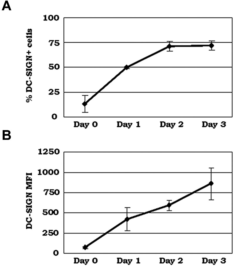
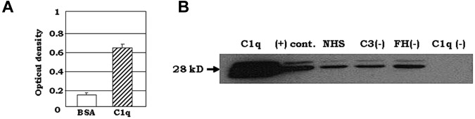
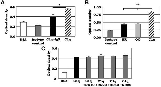
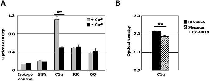
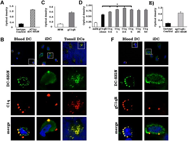
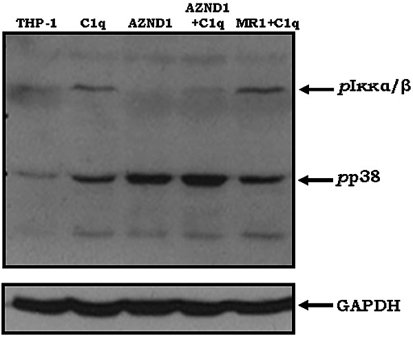
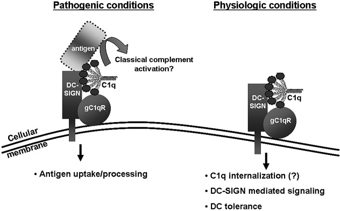
C1q modulates the differentiation and function of cells committed to the monocyte-derived dendritic cell (DC) lineage. Because the 2 C1q receptors found on the DC surface-gC1qR and cC1qR-lack a direct conduit into intracellular elements, we postulated that the receptors must form complexes with transmembrane partners. In the present study, we show that DC-SIGN, a C-type lectin expressed on DCs, binds directly to C1q, as assessed by ELISA, flow cytometry, and immunoprecipitation experiments. Surface plasmon resonance analysis revealed that the interaction was specific, and both intact C1q and the globular portion of C1q bound to DC-SIGN. Whereas IgG reduced this binding significantly, the Arg residues (162-163) of the C1q-A chain, which are thought to contribute to the C1q-IgG interaction, were not required for C1q binding to DC-SIGN. Binding was reduced significantly in the absence of Ca(2+) and by preincubation of DC-SIGN with mannan, suggesting that C1q binds to DC-SIGN at its principal Ca(2+)-binding pocket, which has increased affinity for mannose residues. Antigen-capture ELISA and immunofluorescence microscopy revealed that C1q and gC1qR associate with DC-SIGN on blood DC precursors and immature DCs. The results of the present study suggest that C1q/gC1qR may regulate DC differentiation and function through the DC-SIGN-mediated induction of cell-signaling pathways.


Figures

Figure 1
Figure 1
Surface expression of DC-SIGN increases as monocytes differentiate into iDCs. PBMCs were isolated by density gradient centrifugation and cultured for 3 days in the presence of DC growth factors. Cells were collected on each day and incubated with anti–DC-SIGN mAb, followed by incubation with a secondary Ab conjugated to Alexa Fluor 488 and analysis by flow cytometry. (A) Percentage of positive cells. (B) Mean fluorescence intensity (MFI; n = 3).
Figure 2
Figure 2
C1q binds to DC-SIGN. (A) C1q was added to DC-SIGN–coated plates and binding was detected using pAb anti-C1q Ab in an antigen-capture ELISA. One representative experiment is shown (n = 3). (B) Normal human serum (NHS; 150 μL) that was C3, factor H (FH), and C1q depleted was incubated overnight at 4°C with 2 μg of biotinylated DC-SIGN and added to neutravidin-coated resin beads for 2 hours at RT. Bead-bound proteins were visualized by Western blotting using pAb C1q, followed by HRP- conjugated secondary Ab. Soluble C1q protein was used instead of human serum as a positive control, shown as “(+) cont.” (n = 2).
Figure 3
Figure 3
DC-SIGN binds to C1q at the IgG-binding site. (A) IgG reduced the binding of C1q to DC-SIGN significantly. ELISA experiments were performed using C1q premixed with human IgG in a 1:10 molar ratio for 15 minutes at 37°C. C1q/IgG was added to DC-SIGN– or BSA-coated plates and binding was detected using C1q pAb. One representative experiment is shown (n = 3). *P < .05. (B) Synthetic peptide corresponding to C1q-A chain residues 155-164 bound to DC-SIGN, and the Arg residues (162-163) were not required for binding. ELISA binding studies were performed using 2 synthetic peptides, the C1q-A-chain peptide containing 2 adjacent Arg residues (RR) and another peptide that had 2 glutamines (QQ) substituting for the 2 Arg residues at positions 162-163 (n = 3). *P < .05. (C) Competition of a synthetic peptide corresponding to C1q-A 155-164 did not diminish binding of C1q to DC-SIGN. Competition ELISA experiments were performed using purified, soluble C1q and the synthetic C1q peptide RR. Biotinylated C1q and increasing concentrations of RR (0-80 μg/mL) were added to DC-SIGN–coated plates and binding was detected using anti-C1q oAb. One representative experiment is shown (n = 3).
Figure 4
Figure 4
The C1q–DC-SIGN interaction is Ca2+ dependent and reduced by mannan. (A) ELISA experiments were performed using purified, soluble C1q or various synthetic C1q-A peptides (aa 155-164 for RR, aa 155-164 for RR at 162/163 changed to QQ). Then, 2.5 μg/mL of biotinylated DC-SIGN was added to the C1q- or synthetic peptide–coated plates in the presence or absence of 10mM EDTA and binding was detected using streptavidin-AP. One representative experiment is shown (n = 3). (B) Mannan reduced the binding of C1q to DC-SIGN significantly. ELISA experiments were performed using 2.5 μg/mL of biotinylated DC-SIGN premixed with 2 mg/mL of mannan for 15 minutes at RT. DC-SIGN/mannan was added to C1q- or BSA-coated plates and binding was detected using streptavidin-AP. One representative experiment is shown (n = 3). **P < .01.
Figure 5
Figure 5
DC-SIGN colocalized with C1q and gC1qR on DCs. (A) DC-SIGN was cocaptured with C1q from iDC lysates. Whole-cell lysates (day 3) were added to microtiter plates coated with mAb DC-SIGN and the presence of C1q was detected using anti-C1q pAb. One representative experiment is shown (n = 3). (B) DC-SIGN colocalizes with C1q on the surface of iDCs in vitro, on DC precursors in blood and on iDCs in human tonsils in vivo. PBMCs, day 3 iDCs, and cryostat tonsil sections were analyzed for DC-SIGN and C1q. Cells and tonsil sections were incubated with rat anti–DC-SIGN and goat anti-C1q Abs or isotype controls, followed by FITC–anti–rat and PE-anti–goat secondary Abs and DAPI (blue). The slides were viewed using a Zeiss Axiovert 200M digital deconvolution microscope (63×; oil) and analyzed with AxioVision Version 4.8 software. Isotype controls showed little or no staining (data not shown; n = 6). (C) DC-SIGN binds to gC1qR. Antigen-capture ELISA experiments were performed using purified, soluble gC1qR. gC1qR was added to DC-SIGN–coated plates and binding was detected using anti-gC1qR pAb. One representative experiment is shown (n = 3). (D) C1q increased the binding of gC1qR to DC-SIGN significantly. ELISA experiments were performed using 5 μg/mL of biotinylated gC1qR premixed with increasing concentrations (0-50 μg/mL) of C1q. Biot-gC1qR/C1q was added to DC-SIGN– or 5% nonfat milk–coated plates and binding was detected using neutravidin-AP. One representative experiment is shown (n = 2). *P < .05. (E) DC-SIGN was cocaptured with gC1qR from iDC lysates. Antigen-capture ELISA experiments were performed using whole-cell DC lysates (day 3). DC lysates were added to microtiter plates coated with rat anti–human DC-SIGN and the presence of gC1qR was detected using anti-gC1qR pAb. One representative experiment is shown (n = 3). (F) DC-SIGN colocalizes with gC1qR on the surface of blood DC precursors and iDCs. PBMCs and day 3 iDCs were incubated with rat anti–DC-SIGN and rabbit anti-gC1qR Abs or isotype controls, followed by FITC-anti–rat and PE–anti–rabbit secondary Abs and DAPI (blue). The slides were viewed using a Zeiss Axiovert 200M digital deconvolution microscope (68×; oil) and analyzed with AxioVision Version 4.8 software. Isotype controls showed little or no staining (data not shown; n = 7).
Figure 6
Figure 6
C1q-mediated activation of NF-κB requires DC-SIGN. Phosphorylation of Iκκα/β in the DC-SIGN–expressing human THP-1 cell line was determined after treatment with Abs to DC-SIGN (AZND1 and MR-1; 20 μg/mL at 37°C for 20 minutes) and/or C1q (at 37°C for 10 minutes). After cell lysis, phosphorylated Iκκα/β and p38 were detected using specific mAbs. The blot was then stripped and probed for GAPDH levels as a control for protein loading (n = 2).
Figure 7
Figure 7
Hypothetical model for the gC1qR/C1q–DC-SIGN complex under pathologic and physiologic conditions.

Similar articles

Cited by

References

    1. Castellano G, Woltman AM, Nauta AJ, et al. Maturation of dendritic cells abrogates C1q production in vivo and in vitro. Blood. 2004;103:3813–3820. - PubMed
    1. Kaul M, Loos M. Expression of membrane C1q in human monocyte-derived macrophages is developmentally regulated and enhanced by interferon-gamma. FEBS Lett. 2001;500(1-2):91–98. - PubMed
    1. Reid KB. Chemistry and molecular genetics of C1q. Behring Inst Mitt. 1989;84:8–19. - PubMed
    1. Lu J, Wiedemann H, Timpl R, Reid KB. Similarity in structure between C1q and the collectins as judged by electron microscopy. Behring Inst Mitt. 1993;93:6–16. - PubMed
    1. Bobak DA, Gaither TA, Frank MM, Tenner AJ. Modulation of FcR function by complement: subcomponent C1q enhances the phagocytosis of IgG-opsonized targets by human monocytes and culture-derived macrophages. J Immunol. 1987;138(4):1150–1156. - PubMed

Publication types

MeSH terms