

A comparison of radiation exposure between diagnostic CTA and DSA examinations of cerebral and cervicocerebral vessels
- PMID: 22700752
- PMCID: PMC7965573
- DOI: 10.3174/ajnr.A3123
A comparison of radiation exposure between diagnostic CTA and DSA examinations of cerebral and cervicocerebral vessels
Abstract
Background and purpose: While the number of CTA examinations is continually increasing compared with DSA examinations, there is little comparative dose information about the different imaging techniques. We compared patient radiation exposure resulting from diagnostic CTA and DSA examinations for both cerebral and cervicocerebral vessels.
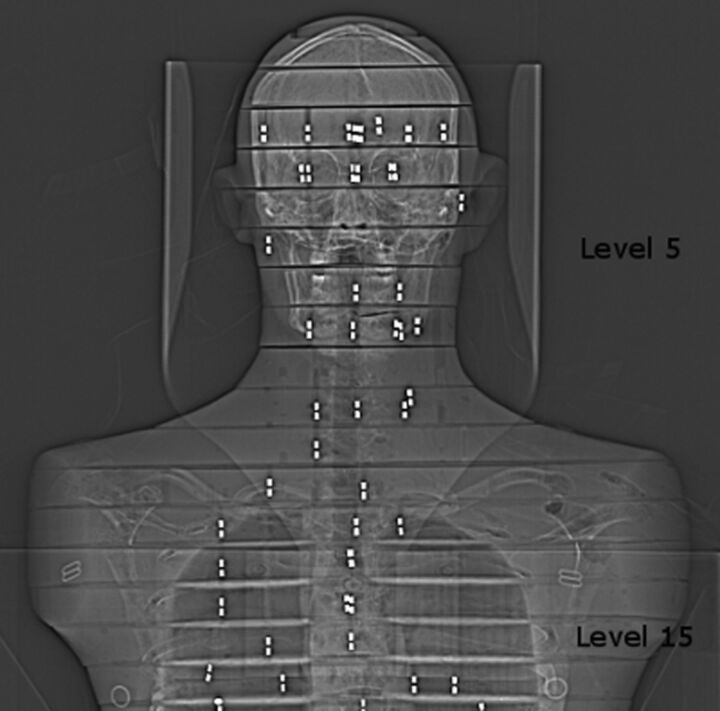
Materials and methods: An anthropomorphic phantom was irradiated by using typical diagnostic CTA and DSA setups and imaging parameters. For both imaging techniques, the imaging area of cerebral vessels included intracranial vessels only, while the imaging area of cervicocerebral vessels included both cervical and intracranial vessels from the aortic arch to the vertex. The effective dose was determined by using RPLDs. The DSA examination was simulated by using a biplane angiography system, and the CTA examination, by using a 64-row multidetector CT scanner.
Results: For the imaging of cerebral vessels, the effective dose according to ICRP 103 was 0.67 mSv for CTA and 2.71 mSv for DSA. For the imaging of cervicocerebral vessels, the effective dose was 4.85 mSv for CTA and 3.60 mSv for DSA. The maximum absorbed dose (milligray) for skin, brain, salivary glands, and eyes was 166.2, 73.5, 35.6, and 21.8 mGy for DSA and 19.0, 16.9, 20.4, and 14.8 mGy for CTA, respectively. The conversion factors from DAP and DLP to effective dose were calculated.
Conclusions: The effective dose for CTA assessment of cerebral vessels was approximately one-fifth the dose compared with DSA. In the imaging of cervicocerebral vessels, the effective dose for CTA was approximately one-third higher compared with DSA.
Figures
References
-
- Teksam M, McKinney A, Cakir B, et al. . Multi-slice CT angiography of small cerebral aneurysms: is the direction of aneurysm important in diagnosis? Eur J Radiol 2005;53:454–62 - PubMed
-
- Bridcut RR, Murphy E, Workman A, et al. . Patient dose from 3D rotational neurovascular studies. Br J Radiol 2007;80:362–66 - PubMed
-
- Schuknecht B. Latest techniques in head and neck CT angiography. Neuroradiology 2004;46:208–13 - PubMed
Publication types
MeSH terms
LinkOut - more resources
Full Text Sources
Medical