Review
. 2012 Aug 1;4(8):a012872.
doi: 10.1101/cshperspect.a012872.

Protein secretion and the endoplasmic reticulum

Affiliations
Review

Protein secretion and the endoplasmic reticulum

Adam M Benham. Cold Spring Harb Perspect Biol. .

Abstract

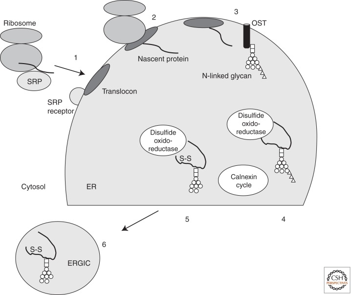
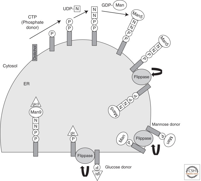
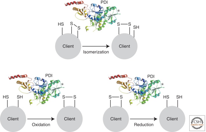
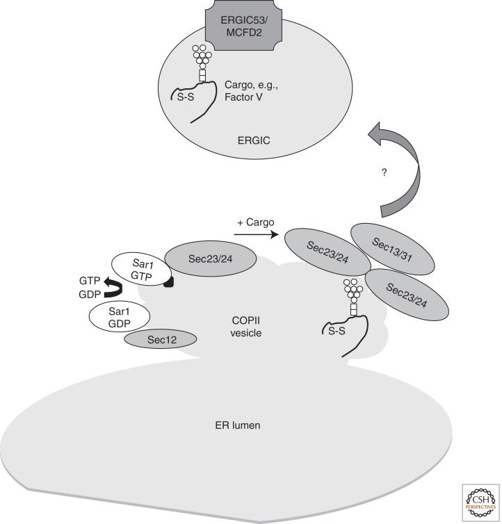
In a complex multicellular organism, different cell types engage in specialist functions, and as a result, the secretory output of cells and tissues varies widely. Whereas some quiescent cell types secrete minor amounts of proteins, tissues like the pancreas, producing insulin and other hormones, and mature B cells, producing antibodies, place a great demand on their endoplasmic reticulum (ER). Our understanding of how protein secretion in general is controlled in the ER is now quite sophisticated. However, there remain gaps in our knowledge, particularly when applying insight gained from model systems to the more complex situations found in vivo. This article describes recent advances in our understanding of the ER and its role in preparing proteins for secretion, with an emphasis on glycoprotein quality control and pathways of disulfide bond formation.


Figures

Figure 1.
Figure 1.
Overview of protein targeting to the ER and glycoprotein quality control. Six key points in the fidelity of protein secretion are illustrated. (1) Correct targeting of the glycoprotein to the ER as it emerges from the translocon. This is mediated by the signal recognition particle (SRP) and its receptor, which helps position the emerging protein at the translocon. (2) Translocation of the (glyco)protein into the ER by the translocon. (3) Addition of N-glycans to Asn residues of glycoproteins by oligosaccharyl transferase (OST). (4) Correct folding and quality control by the calnexin cycle. (5) Introduction/correction of disulfide bonds by protein disulfide isomerases/oxidoreductases. (6) Directing glycoproteins into ER exit sites followed by packaging into appropriate compartments, for example, the ER Golgi intermediate compartment (ERGIC). Note that protein folding and disulfide bond formation happen rapidly after the nascent protein emerges from the translocon. (S–S) Disulfide bond; (□) N-Acetyl glucosamine; (○) mannose; (▵) glucose.
Figure 2.
Figure 2.
The N-linked glycosylation pathway. N-linked glycosylation begins in the cytosol. Phosphate (P) from CTP is used to charge dolichol in the lipid bilayer. Dolichol phosphate receives two N-acetyl glucosamine moieties (N) from UDP-N-acetyl glucosamine. Five GDP-mannose (Man) residues are added sequentially before the sugar donor is translocated across the ER membrane by the “flippase.” In the ER lumen, four additional mannose and three glucose residues are added to yield a dolichol-linked Glc(3)Man(9)GlcNAc(2) structure that is transferred to an Asn residue of a nascent glycoprotein by OST. The identity of the “flippases” that translocate Man(5)GlcNAc(2)-P-P-dolichol, Man-P-dolichol, and Glc-P-dolichol are debated (see text for details). (□) N-Acetyl glucosamine; (○) mannose; (▵) glucose.
Figure 3.
Figure 3.
Disulfide bond formation in the ER. Disulfide bonds form between the –SH groups of two cysteine residues in a protein. The process is mostly confined to the ER in eukaryotes and is catalyzed by enzymes. The most abundant catalyst of disulfide bond formation is PDI, which can introduce (oxidize), remove (reduce), or swap (isomerize) disulfide bonds in a range of client proteins.
Figure 4.
Figure 4.
ER exit, COPII vesicles, and ERGIC. COPII exit vesicles form when Sec12p recruits Sar1p-GDP and exchanges GDP for GTP, enabling Sar1p to insert into the budding membrane. Sar1p facilitates the assembly of Sec23/24 and Sec13/31 at the membrane upon recruitment of cargo. The relationship between COPII vesicles and the ER Golgi intermediate compartment (ERGIC) is not comprehensively defined in higher eukaryotes, and some cargoes may escape the ER directly by bulk flow. ERGIC53 and MCFD2 are required to recruit at least some cargoes to the ERGIC (e.g., Factor V and VIII). (□) N-Acetyl glucosamine; (○) mannose.

References

    1. Acar M, Jafar-Nejad H, Takeuchi H, Rajan A, Ibrani D, Rana NA, Pan H, Haltiwanger RS, Bellen HJ 2008. Rumi is a CAP10 domain glycosyltransferase that modifies Notch and is required for Notch signaling. Cell 132: 247–258 - PMC - PubMed
    1. Alder NN, Shen Y, Brodsky JL, Hendershot LM, Johnson AE 2005. The molecular mechanisms underlying BiP-mediated gating of the Sec61 translocon of the endoplasmic reticulum. J Cell Biol 168: 389–399 - PMC - PubMed
    1. Anelli T, Ceppi S, Bergamelli L, Cortini M, Masciarelli S, Valetti C, Sitia R 2007. Sequential steps and checkpoints in the early exocytic compartment during secretory IgM biogenesis. EMBO J 26: 4177–4188 - PMC - PubMed
    1. Anelli T, Bergamelli L, Margittai E, Rimessi A, Fagioli C, Malgaroli A, Pinton P, Ripamonti M, Rizzuto R, Sitia R 2012. Ero1α regulates Ca2+ fluxes at the endoplasmic reticulum-mitochondria interface (MAM). Antioxid Redox Signal 16: 1077–1087 - PubMed
    1. Appenzeller-Herzog C 2011. Glutathione- and non-glutathione-based oxidant control in the endoplasmic reticulum. J Cell Sci 124: 847–855 - PubMed

Publication types

LinkOut - more resources