Comparative Study
. 2012 Aug;110(3):595-613.
doi: 10.1093/aob/mcs119. Epub 2012 Jun 13.

A survey of mangiferin and hydroxycinnamic acid ester accumulation in coffee (Coffea) leaves: biological implications and uses

Affiliations
Comparative Study

A survey of mangiferin and hydroxycinnamic acid ester accumulation in coffee (Coffea) leaves: biological implications and uses

Claudine Campa et al. Ann Bot. 2012 Aug.

Abstract

Background and aims: The phenolic composition of Coffea leaves has barely been studied, and therefore this study conducts the first detailed survey, focusing on mangiferin and hydroxycinnamic acid esters (HCEs).

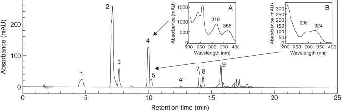
Methods: Using HPLC, including a new technique allowing quantification of feruloylquinic acid together with mangiferin, and histochemical methods, mangiferin content and tissue localization were compared in leaves and fruits of C. pseudozanguebariae, C. arabica and C. canephora. The HCE and mangiferin content of leaves was evaluated for 23 species native to Africa or Madagascar. Using various statistical methods, data were assessed in relation to distribution, ecology, phylogeny and use.

Key results: Seven of the 23 species accumulated mangiferin in their leaves. Mangiferin leaf-accumulating species also contain mangiferin in the fruits, but only in the outer (sporophytic) parts. In both leaves and fruit, mangiferin accumulation decreases with ageing. A relationship between mangiferin accumulation and UV levels is posited, owing to localization with photosynthetic tissues, and systematic distribution in high altitude clades and species with high altitude representatives. Analyses of mangiferin and HCE content showed that there are significant differences between species, and that samples can be grouped into species, with few exceptions. These data also provide independent support for various Coffea lineages, as proposed by molecular phylogenetic analyses. Sampling of the hybrids C. arabica and C. heterocalyx cf. indicates that mangiferin and HCE accumulation may be under independent parental influence.

Conclusions: This survey of the phenolic composition in Coffea leaves shows that mangiferin and HCE accumulation corresponds to lineage recognition and species delimitation, respectively. Knowledge of the spectrum of phenolic accumulation within species and populations could be of considerable significance for adaptation to specific environments. The potential health benefits of coffee-leaf tea, and beverages and masticatory products made from the fleshy parts of Coffea fruits, are supported by our phenolic quantification.


Figures

Fig. 1.
Fig. 1.
HPLC profile of a C. pseudozanguebariae leaf extract. Absorption profile obtained at 320 nm using the technical conditions for quantitative analysis. Peak 1 = 3-caffeoylquinic acid (3-CQA); peak 2 = 5-caffeoylquinic acid (5-CQA); peak 3 = 4-caffeoylquinic acid (4-CQA); peak 4 = mangiferin; peak 4' = isomangiferin; peak 5 = 5-feruloylquinic acid (5-FQA); peak 6 = 4-feruloylquinic acid (4-FQA); peak 7 = 3,4-dicaffeoylquinic acid (3,4 diCQA); peak 8 = 3,5-dicaffeoylquinic acid (3,5-diCQA); and peak 9 = 4,5-dicaffeoylquinic acid (4,5-diCQA). The absorption spectrum between 200 and 400 nm is indicated for mangiferin (A) and FQA (B).
Fig. 2.
Fig. 2.
Histochemical localization of mangiferin in leaves (A) and green fruit (B) of different Coffea species. Cross-sections were observed under UV light, ×400, except B4 (×40). (A) Leaves. Coffea pseudozanguebariae leaf blade (A1), showing a very high concentration of mangiferin preferentially localized in the cells forming the palisade and spongy parenchyma (mesophyll) (yellow arrows). Coffea canephora leaf blade (A2), no mangiferin accumulated (i.e. no specific yellow autofluorescence), allowing observation of chlorophyll (red autofluorescence), principally in palisade and spongy parenchyma. Blue fluorescences are specific for cuticle constituents and of caffeoylquinic acids (in epidermal cells). Coffea arabica ‘Laurina’ leaf blade (A3), showing that the presence of mangiferin (yellow autofluorescence) has attenuated the red autofluorescence of the chlorophyll. (B) Fruits. Coffea pseudozanguebariae (B1), yellow autofluorescence of transverse sections (T.S.) of immature fruits (stage 1), showing that mangiferin is extremely concentrated in the exocarp and mesocarp cells. Coffea canephora (B2), no mangiferin accumulated. Coffea arabica ‘Laurina’ (B3 and B4) mangiferin accumulation appears to be vacuolar and restricted to exocarp and external mesocarp cells (arrows) in young fruits. There is no mangiferin detected in the endocarp, integument (seed coat) and seed. Abbreviations: c, cuticle; chl, chlorophyll; e, epidermis; en, endocarp; ex, exocarp; in, integument; m, mesocarp; pp, palisade; s, seed; sp, spongy parenchyma (mesophyll).
Fig. 3.
Fig. 3.
Linear regression between 4-CQA and 3-CQA contents in leaves. Analysis included 40 genotypes from the 15 African taxa.
Fig. 4.
Fig. 4.
Hierarchical clustering analyses of African Coffea taxa using seven HCE variables (see Table 5). (A) Taxa from the Upper and Lower Guinea/Congolian regions (White, 1979, 1983), and the hybrids C. arabica ‘Laurina’ and C. heterocalyx cf. (B) Taxa from East Central Africa and East Africa. (see Tables 1, 5 and 6, and text, for details).
Fig. 5.
Fig. 5.
Principal component analysis (PCA) using seven HCE variables (see Table 5). (A) Projection of the seven variables on the axes: 3-CQA, 4-CQA, 5-CQA, 3,4 diCQA, 3,5 diCQA, 4,5 diCQA and FQAs. (B) Score plot of the 40 African genotypes. (C) Enlargement of the central zone of the score plot. Grouped genotypes from individualized species are noted in the text. Abbreviations: ant, C. anthonyi; can 1, C. canephora (Congo); can 2, C. canephora (Cameroon); con, C. congensis; eug, C. eugenioides; het, C. heterocalyx cf.; hum, C. humilis; lib, C. liberica var. liberica; dew, C. liberica var. dewevrei; man, C. mannii; pseu, C. pseudozanguebariae; rac, C. racemosa; sal, C. salvatrix; ses, C. sessiliflora; sten, C. stenophylla; arab, C. arabica ‘Laurina’.

References

    1. Abd El-Mawla AMA, Beerhues L. Benzoic acid biosynthesis in cell cultures of Hypericum androsaemum. Planta. 2002;214:727–733. - PubMed
    1. Alonso-Salces RM, Guillou C, Berrueta LA. Liquid chromatography coupled with ultraviolet absorbance detection, electrospray ionization, collision-induced dissociation and tandem mass spectrometry on a triple quadrupole for the on-line characterization of polyphenols and methylxanthines in green coffee beans. Rapid Communications in Mass Spectrometry. 2009;23:363–383. - PubMed
    1. Anonymous. Coffee-leaf tea. British Medical Journal. 1876;2(830):691.
    1. Anthony F, Diniz LEC, Combes M-C, Lashermes P. Adaptive radiation in Coffea subgenus Coffea L. (Rubiaceae) in Africa and Madagascar. Plant Systematics and Evolution. 2010;285:51–64.
    1. Anthony F, Noirot M, Clifford MN. Biochemical diversity in the genus Coffea L.: chlorogenic acids, caffeine, and mozambioside contents. Genetic Resources and Crop Evolution. 1993;40:61–70.

Publication types