Clinical Trial
. 2012 Jun 13;4(138):138ra77.
doi: 10.1126/scitranslmed.3003578.

Cell carriage, delivery, and selective replication of an oncolytic virus in tumor in patients

Affiliations
Clinical Trial

Cell carriage, delivery, and selective replication of an oncolytic virus in tumor in patients

Robert A Adair et al. Sci Transl Med. .

Abstract

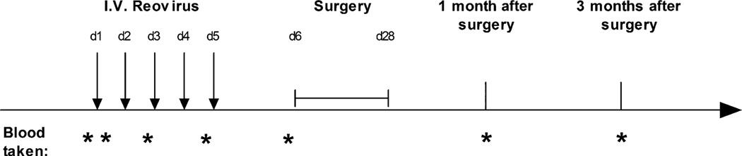
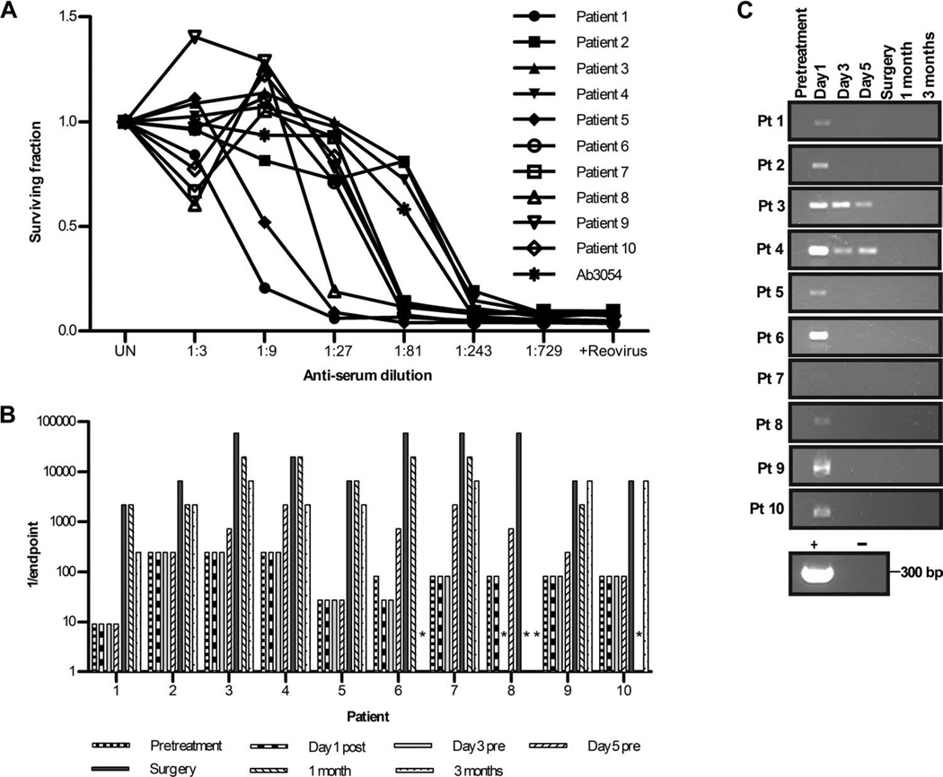
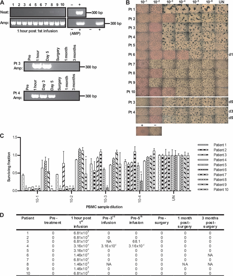
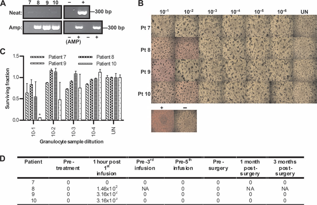
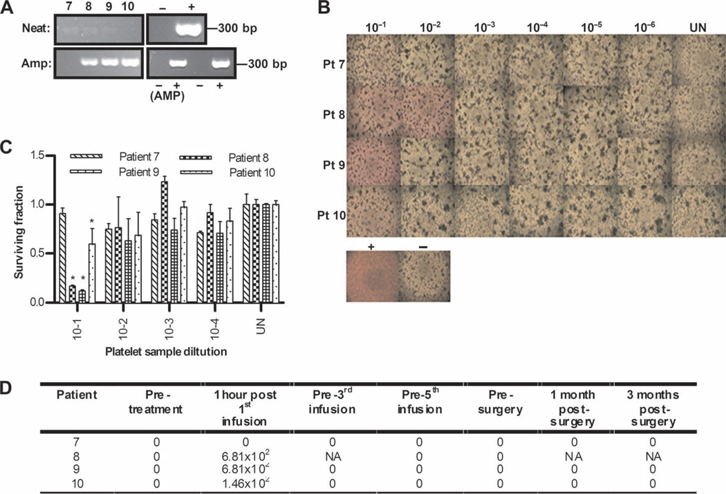
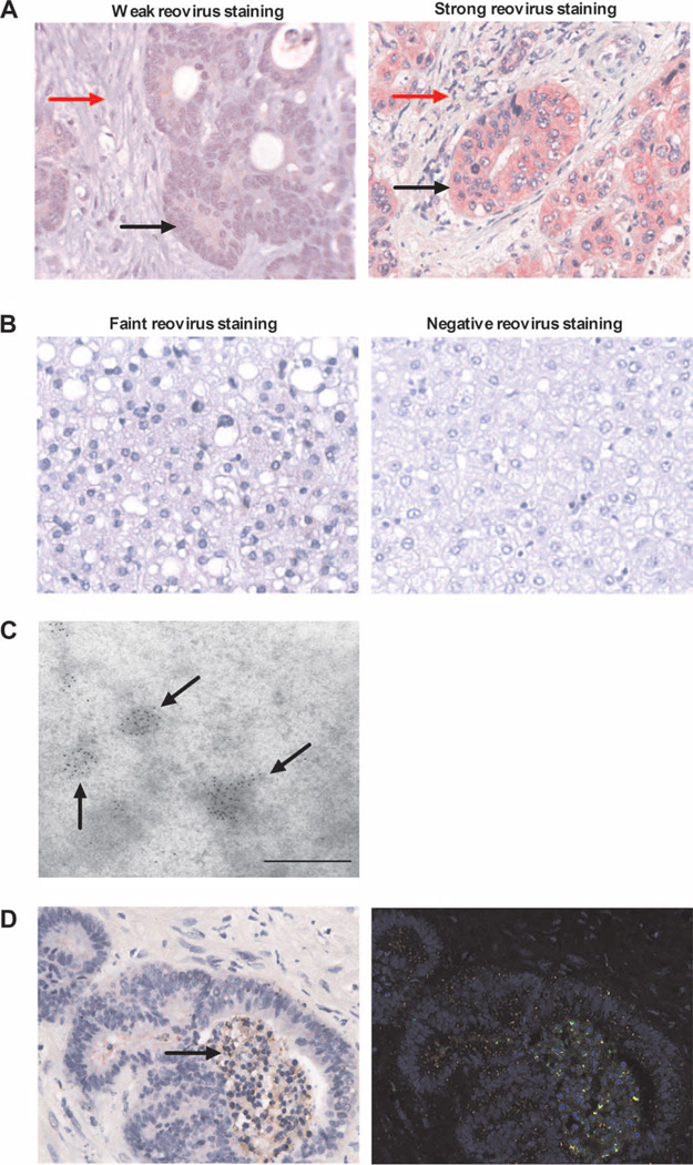
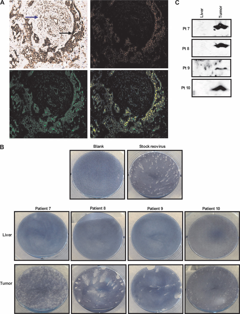
Oncolytic viruses, which preferentially lyse cancer cells and stimulate an antitumor immune response, represent a promising approach to the treatment of cancer. However, how they evade the antiviral immune response and their selective delivery to, and replication in, tumor over normal tissue has not been investigated in humans. Here, we treated patients with a single cycle of intravenous reovirus before planned surgery to resect colorectal cancer metastases in the liver. Tracking the viral genome in the circulation showed that reovirus could be detected in plasma and blood mononuclear, granulocyte, and platelet cell compartments after infusion. Despite the presence of neutralizing antibodies before viral infusion in all patients, replication-competent reovirus that retained cytotoxicity was recovered from blood cells but not plasma, suggesting that transport by cells could protect virus for potential delivery to tumors. Analysis of surgical specimens demonstrated greater, preferential expression of reovirus protein in malignant cells compared to either tumor stroma or surrounding normal liver tissue. There was evidence of viral factories within tumor, and recovery of replicating virus from tumor (but not normal liver) was achieved in all four patients from whom fresh tissue was available. Hence, reovirus could be protected from neutralizing antibodies after systemic administration by immune cell carriage, which delivered reovirus to tumor. These findings suggest new preclinical and clinical scheduling and treatment combination strategies to enhance in vivo immune evasion and effective intravenous delivery of oncolytic viruses to patients in vivo.


Figures

Fig. 1
Fig. 1
Trial schema is presented: timing of reovirus infusion, sample collection, and surgery. I.V., intravenous.
Fig. 2
Fig. 2
NABs are present at baseline and increase after treatment, whereas the viral genome is only transiently detectable after infusion in plasma. (A) Samples collected before virus infusion were tested for baseline NAB levels. Plot shows neutralization of reovirus-induced killing of L929 cells by serial dilutions of samples as measured by MTT assay at 72 hours. L929 cells were treated with reovirus only (+Reovirus) or left untreated (UN) as positive and negative controls, respectively; anti-reovirus polyclonal goat antibody (Ab3054) was used as a standard curve. (B) Changes in endpoint reovirus NAB titer over time (asterisk denotes samples unavailable for analysis). (C) Patient plasma was assessed for reovirus RNA by RT-PCR over time. Reovirus RNA and RNase-free water were included as positive and negative controls, respectively. bp, base pairs.
Fig. 3
Fig. 3
Despite circulating NABs, PBMCs transiently carry reovirus after infusion, which can replicate in and kill target cells in vitro. (A) Day 1 post-infusion PBMCs were assessed directly for reovirus RNA by RT-PCR (neat) or after an additional amplification step on L929 cells for 7 days (amplified). Reovirus RNA and RNase-free water were included as positive and negative controls, respectively, alongside a 1:10 dilution of stock reovirus or 5% DMEM incubated on L929 cells as amplified positive and negative controls (AMP). Later time points for patients 3 and 4 are also shown. (B) PBMCs were assessed for functional reovirus in a TCID50 assay. L929 cells were cultured with serial dilutions of PBMCs and observed 7 days later for CPE. Photomicrographs show results from day 1 post-infusion PBMCs for all samples and later time points for patients 3 and 4. Dilution (1:10) of stock reovirus or 5% DMEM (UN) was incubated on L929 cells as positive and negative controls, respectively. Rounded up cells and unused (red) media signify CPE. (C) Reovirus-induced cell killing by day 1 posttreatment PBMCs was further confirmed by MTT analysis. *P < 0.05 versus untreated control; error bars represent SEM. (D) Viral titers (TCID50/ml) from PBMCs over time (NA denotes samples unavailable for analysis).
Fig. 4
Fig. 4
Granulocytes similarly carry replication-competent reovirus after infusion. (A) Day 1 post-infusion granulocytes were assessed for reovirus RNA by RT-PCR, using both neat and amplified samples as for PBMCs in Fig. 3A. (B) Granulocytes were assessed for functional reovirus in a TCID50 assay as for PBMCs in Fig. 3B. Photomicrographs show day 1 post-infusion granulocyte dilutions; rounded up cells and unused (red) media signify CPE. (C) Reovirus-induced cell killing by day 1 posttreatment granulocytes was further confirmed by MTT analysis. *P < 0.05 versus untreated control; error bars represent SEM. (D) Viral titers (TCID50/ml) from granulocytes over time (NA denotes samples unavailable for analysis).
Fig. 5
Fig. 5
Platelets also carry reovirus after infusion. (A) Day 1 post-infusion platelets were assessed for reovirus RNA by RT-PCR, using both neat and amplified samples as for PBMCs in Fig. 3A. (B) Platelets were assessed for functional reovirus in a TCID50 assay as for PBMCs in Fig. 3B. Photomicrographs show day 1 post-infusion platelet dilutions; rounded up cells and unused (red) media signify CPE. (C) Reovirus-induced cell killing by day 1 posttreatment platelets was further confirmed by MTT analysis. *P < 0.05 versus untreated control; error bars represent SEM. (D) Viral titers (TCID50/ml) in platelets over time (NA denotes samples unavailable for analysis).
Fig. 6
Fig. 6
Intravenous reovirus is preferentially detected in metastatic colorectal tumor cells within the liver. (A) Immunohistochemistry images showing expression of reovirus protein (red stain) in resected colorectal liver metastases (magnification, ×400). One representative case each of weak (left; patient 1) and strong (right; patient 6) staining is shown. Malignant cells and tumor stroma are marked by black and red arrows, respectively. (B) Immunohistochemistry images for expression of reovirus protein (red stain) in normal liver (magnification, ×400). One representative case each of faint (left; patient 8) and negative (right; patient 2) staining is shown. (C) Representative EM image (patient 8) showing immunogold staining of reovirus σ3 capsid protein within colorectal liver metastases. Scale bar, 500 nm. (D) RGB image analyses of resected colorectal liver metastases, using the Nuance System (magnification, ×400). Images are representative (patient 9) and show reovirus staining (red) and caspase-3 staining (brown) (left image; arrow indicates changes of nuclear and cytoplasmic degeneration in reovirus-infected tumor cells). Right image shows conversion of RGB image to fluorescent green (caspase-3), fluorescent red (reovirus), and yellow (coexpression).
Fig. 7
Fig. 7
Replication-competent reovirus can be retrieved from tumor tissue. (A) RGB image analyses of resected colorectal liver metastases, using the Nuance System (magnification, ×200). Images are representative (patient 10) and show (top left) reovirus staining (red) and tubulin staining (brown) (malignant cells and tumor stroma are marked by black and blue arrows, respectively), (top right) conversion of RGB image to fluorescent red (reovirus), (bottom left) conversion of RGB image to fluorescent green (tubulin), and (bottom right) coexpression of reovirus and tubulin (yellow). (B) Plaque assays demonstrating retrieval of reovirus from freshly resected tumor and liver tissue; photographs show representative wells of optimized supernatant dilutions of 1:2500 (patient 7), neat supernatant (patients 8 and 9), and 1:1200 (patient 10). Photographs of all liver samples show representative wells of neat supernatant. (C) Culture supernatants from plaque assays performed in (B) were assessed for reovirus σ3 capsid protein by Western blotting.

References

    1. Garcia-Aragoncillo E, Hernandez-Alcoceba R. Design of virotherapy for effective tumor treatment. Curr. Opin. Mol. Ther. 2010;12:403–411. - PubMed
    1. Prestwich RJ, Harrington KJ, Pandha HS, Vile RG, Melcher AA, Errington F. Oncolytic viruses: A novel form of immunotherapy. Expert Rev. Anticancer Ther. 2008;8:1581–1588. - PMC - PubMed
    1. Eager RM, Nemunaitis J. Clinical development directions in oncolytic viral therapy. Cancer Gene Ther. 2011;18:305–317. - PubMed
    1. Kaufman HL, Bines SD. OPTIM trial: A phase III trial of an oncolytic herpes virus encoding GM-CSF for unresectable stage III or IV melanoma. Future Oncol. 2010;6:941–949. - PubMed
    1. Breitbach CJ, Thorne SH, Bell JC, Kirn DH. Targeted and armed oncolytic poxviruses for cancer: The lead example of JX-594. Curr. Pharm. Biotechnol. 2011 - PubMed

Publication types

Substances