Clinical Trial
. 2012 Aug 17;287(34):28975-85.
doi: 10.1074/jbc.M112.372094. Epub 2012 Jun 14.

Mutations in the GlyT2 gene (SLC6A5) are a second major cause of startle disease

Affiliations
Clinical Trial

Mutations in the GlyT2 gene (SLC6A5) are a second major cause of startle disease

Eloisa Carta et al. J Biol Chem. .

Abstract

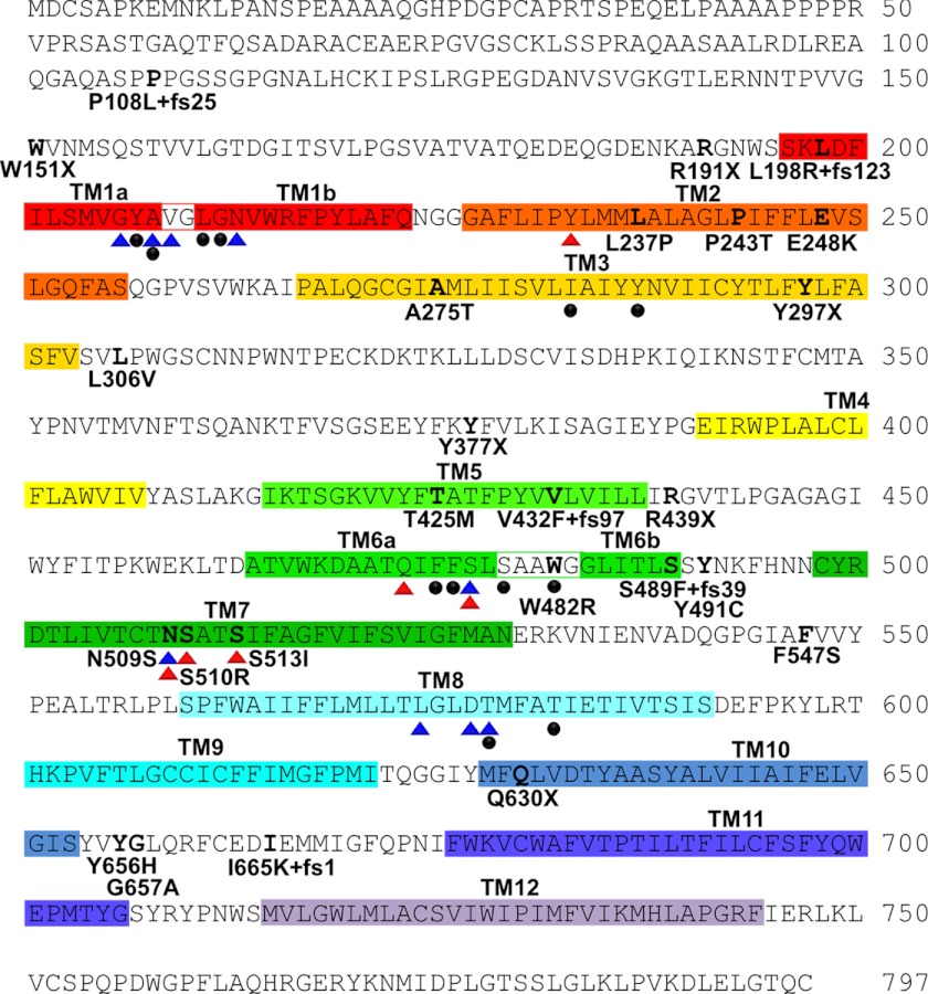
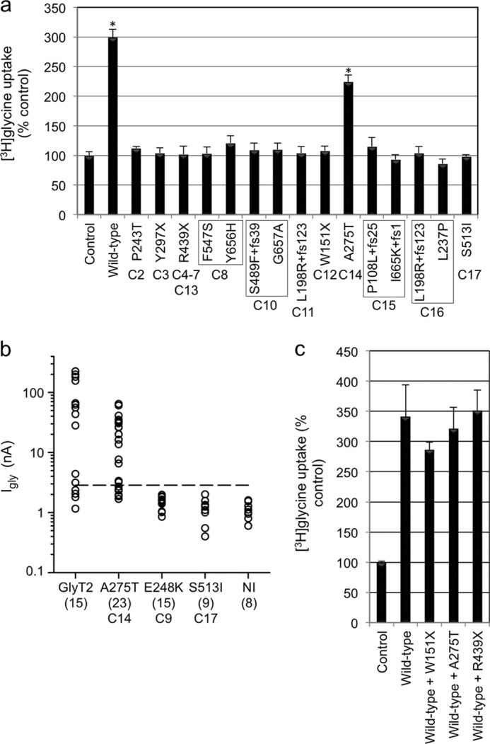
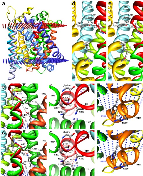
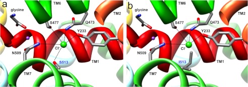
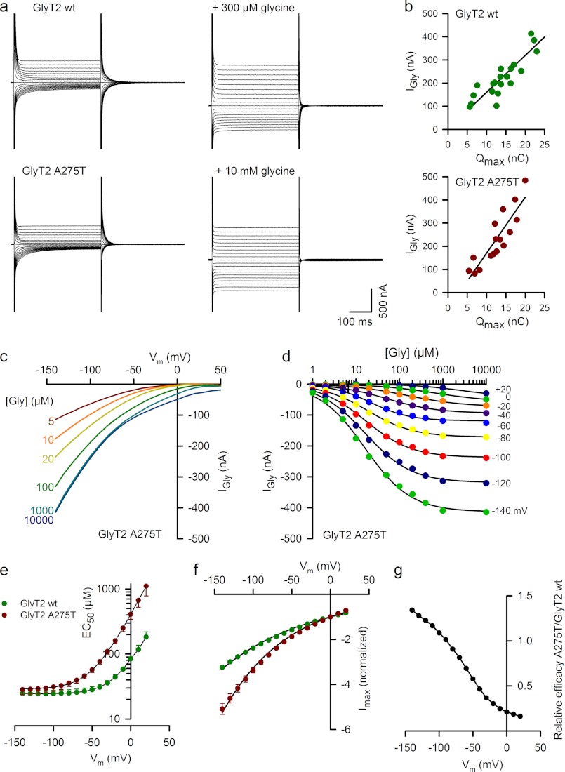
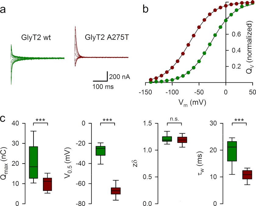
Hereditary hyperekplexia or startle disease is characterized by an exaggerated startle response, evoked by tactile or auditory stimuli, leading to hypertonia and apnea episodes. Missense, nonsense, frameshift, splice site mutations, and large deletions in the human glycine receptor α1 subunit gene (GLRA1) are the major known cause of this disorder. However, mutations are also found in the genes encoding the glycine receptor β subunit (GLRB) and the presynaptic Na(+)/Cl(-)-dependent glycine transporter GlyT2 (SLC6A5). In this study, systematic DNA sequencing of SLC6A5 in 93 new unrelated human hyperekplexia patients revealed 20 sequence variants in 17 index cases presenting with homozygous or compound heterozygous recessive inheritance. Five apparently unrelated cases had the truncating mutation R439X. Genotype-phenotype analysis revealed a high rate of neonatal apneas and learning difficulties associated with SLC6A5 mutations. From the 20 SLC6A5 sequence variants, we investigated glycine uptake for 16 novel mutations, confirming that all were defective in glycine transport. Although the most common mechanism of disrupting GlyT2 function is protein truncation, new pathogenic mechanisms included splice site mutations and missense mutations affecting residues implicated in Cl(-) binding, conformational changes mediated by extracellular loop 4, and cation-π interactions. Detailed electrophysiology of mutation A275T revealed that this substitution results in a voltage-sensitive decrease in glycine transport caused by lower Na(+) affinity. This study firmly establishes the combination of missense, nonsense, frameshift, and splice site mutations in the GlyT2 gene as the second major cause of startle disease.


Figures

FIGURE 1.
FIGURE 1.
Human GlyT2 mutations identified in individuals with startle disease. The amino acid sequence of human GlyT2 indicating the positions of putative transmembrane (TM) domains (colored boxes) and amino acid residues affected by hyperekplexia mutations; this study and Refs. –22). Blue and red triangles indicate residues in hGlyT2 that are likely to coordinate Na+ and Cl ions, respectively, based on structure/function studies of the bacterial leucine transporter LeuT and other mammalian neurotransmitter transporters such as GAT-1 and SERT. However, GlyT2 binds three Na+ ions, whereas LeuT binds two, suggesting that other residues involved in Na+ coordination remain to be identified. Filled black circles indicate residues predicted to be involved in glycine binding.
FIGURE 2.
FIGURE 2.
Functional activity of hGlyT2 and hyperekplexia mutants. a, glycine uptake in HEK293 cells transiently expressing hGlyT2 and single hyperekplexia mutants after 5 min of incubation with [3H]glycine (60 Ci/mmol; PerkinElmer Life Sciences) at a final concentration of 300 μm. Because low levels of glycine uptake are found in HEK293 cells (17), [3H]glycine uptake was calculated as nmol/min/mg of protein and then expressed as a percentage of the empty expression vector transfected control. Only wild-type GlyT2 and mutant A275T result in any detectable uptake above control values. b, steady-state amplitude of the current evoked by 1 mm glycine at −70 mV in Xenopus oocytes injected with selected GlyT2 expression cDNAs. Because 1 mm glycine evokes a small inward current of 1.1 ± 0.34 nA (n = 8) in non-injected (NI) oocytes (because of endogenous amino acid transporters), the threshold for heterologous expression of GlyT2 was set at +5 S.D. (∼2.8 nA, dashed line). Robust currents were observed for 10 of 14 oocytes injected with wild-type GlyT2 and 12 of 18 oocytes injected with the A275T mutant. By contrast, neither E248K or S513I mutants exhibited currents above this threshold (n = 15 and 9, respectively). c, selected [3H]glycine uptake experiments for mutations W151X, A275T, and R439X. We noted no significant effect of co-expressing these mutants with wild-type GlyT2, suggesting that these mutations do not show dominant-negative effects. The data are the means ± S.E. (n = 4–12). Statistical comparisons were made using an unpaired Students t test. For a, the asterisk indicates significantly different from empty vector control. p < 0.01.
FIGURE 3.
FIGURE 3.
Molecular modeling of GlyT2 mutations. a, side view of the human GlyT2 monomer showing transmembrane helices as colored ribbons. Glycine and two (of three) sodium ions (purple spheres) are depicted. Note that the TM3-TM4 extracellular loop (EL2) was not modeled because of an insertion of residues 312–354 in GlyT2 relative to LeuTAA. b and c, substitution E248K in TM2 replaces a negatively charged glutamate for a positively charged lysine, with a longer side chain that introduces potential clashes (red lines) with Ala-480 and Ala-481 in TM6. This is likely to affect the orientation of the key glycine-binding residue Trp-482. d and e, substitution A275T in TM3 results in several predicted clashes with Ile-279 that are predicted to alter the orientation of the potential glycine-binding residue Tyr-287 and destabilize the unwound region of TM1, which contains several determinants of glycine and Na+ binding. f and g, substitution S513I in TM7 introduces a larger side chain at this position, resulting in predicted clashes with Asn-213 (TM1) and Asn-509 (TM7), both of which are involved in the binding of Na+. Note that Asn-509 and Ser-513 are also predicted to be involved in coordinating Cl binding to GlyT2. h and i, substitution Y656H in the intracellular TM10-TM11 loop is predicted to abolish a cation-π interaction that exists between Tyr-656 and Arg-660 that may affect folding or stability or interfere with intracellular GlyT2 accessory protein interactions.
FIGURE 4.
FIGURE 4.
Molecular modeling of the GlyT2 chloride ion-binding site. a, exploded view of the human GlyT2 chloride ion binding site showing Cl as a green sphere and coordination by Tyr-233 in TM2, Gln-473 and Ser-477 in TM6, and Asn-509 and Ser-513 in TM7. b, substitution S513I in TM7 introduces a larger side chain, which lacks the essential hydroxyl necessary for the coordination of Cl and may occlude the binding site. Because a hydroxyl at this position is highly conserved in SLC6 transporters (–29), this substitution is likely to disrupt glycine uptake by directly interfering with coordination of Cl.
FIGURE 5.
FIGURE 5.
Current-voltage relationships and dose-response curves for the GlyT2 mutant A275T. a, current traces evoked by 500-ms voltage steps from +50 mV to −140 mV by decrements of 10 mV in representative oocytes expressing wild-type GlyT2 (upper panels) and the GlyT2 A275T mutant (lower panels) in control solution (left traces) or in the presence of glycine (right traces). b, plots of current amplitude evoked by glycine (1 mm) at −40 mV and Qmax (see “Experimental Procedures” and Fig. 6) relationship for oocytes expressing wild-type GlyT2 (upper panel) or GlyT2 A275T (lower panel). The solid lines show the linear regression with a slope of 15.9 s−1 (r2 = 0.773, n = 20) and 24.1 s−1 (r2 = 0.774, n = 14) for wild-type GlyT2 and A275T, respectively. c, voltage dependence of steady-state currents evoked by the glycine concentrations indicated at left in a representative oocyte expressing GlyT2 A275T. d, dose-response curves of the glycine-evoked current at the membrane potentials indicated on right in the same oocytes as in c. The solid lines are the least square fit of the data to the Hill equation: Igly = Imax/(1 + EC50/[gly])n. e, effect of membrane potential on glycine EC50 for oocytes expressing GlyT2 A275T (red circles, n = 8) and wild-type GlyT2 (green circles, n = 7). The solid lines correspond to the fit of the data to the equation EC50 = A + BαuVm, where A = 23.7 and 29 μm, B = 62.1 and 379 μm, and α = 1.4 and 1.34 for wild-type GlyT2 and A275T, respectively, where u = F/RT (R is the universal gas constant, T is the absolute temperature, and F is Faraday's constant). f, voltage dependence of Imax for wild-type GlyT2 (n = 8) and A275T (n = 7). Imax values were normalized by the values at 0 mV. The solid lines correspond to the fit of the data to the equation Imax = −e −βuVm, with β = 0.3 and 0.22 for wild-type GlyT2 and A275T, respectively. g, relative efficacy (Imax/EC50) of A275T/wild-type GlyT2.
FIGURE 6.
FIGURE 6.
Pre-steady-state kinetics of the GlyT2 mutant A275T. a, relaxation currents recorded in representative oocytes expressing wild-type GlyT2 (green) or GlyT2 A275T (red) during the repolarization to the holding potential (−40 mV) after 400-ms step pulses from +50 to −140 mV. Currents recorded in the presence of ORG25543 were subtracted. b, time integral of the relaxation currents for oocytes expressing wild-type GlyT2 (green circles, n = 14) or GlyT2-A275T (red circles, n = 19). The solid lines are the least square fit of the data to the Boltzmann equation: (QVQmin)/Qmax = 1/(1 + ezδu(V0.5−Vm)) c, box plots of Qmax, V0.5, zδ, and τ weighted distributions for oocytes expressing wild-type GlyT2 (green, n = 14) or GlyT2 A275T (red, n = 19). The box edges represent the 25th and 75th percentiles with the median line. The errors bars represent the 10th and 90th percentiles.

References

    1. Nigro M. A., Lim H. C. (1992) Hyperekplexia and sudden neonatal death. Pediatr. Neurol. 8, 221–225 - PubMed
    1. Vigevano F., Di Capua M., Dalla Bernardina B. (1989) Startle disease. An avoidable cause of sudden infant death. Lancet 1, 216. - PubMed
    1. Bakker M. J., van Dijk J. G., van den Maagdenberg A. M., Tijssen M. A. (2006) Startle syndromes. Lancet Neurol. 5, 513–524 - PubMed
    1. Thomas R. H., Stephenson J. B., Harvey R. J., Rees M. I. (2010) Hyperekplexia. Stiffness, startle and syncope. J. Ped. Neurol. 8, 11–14
    1. Bakker M. J., Peeters E. A., Tijssen M. A. (2009) Clonazepam is an effective treatment for hyperekplexia due to a SLC6A5 (GlyT2) mutation. Mov. Disord. 24, 1852–1854 - PubMed

Publication types

MeSH terms