. 2012 Aug 3;287(32):26854-66.
doi: 10.1074/jbc.M112.363655. Epub 2012 Jun 14.

Structural basis for the interaction of a hexameric replicative helicase with the regulatory subunit of human DNA polymerase α-primase

Affiliations

Structural basis for the interaction of a hexameric replicative helicase with the regulatory subunit of human DNA polymerase α-primase

Bo Zhou et al. J Biol Chem. .

Abstract

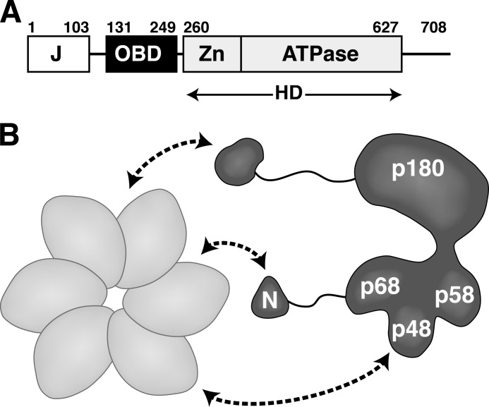
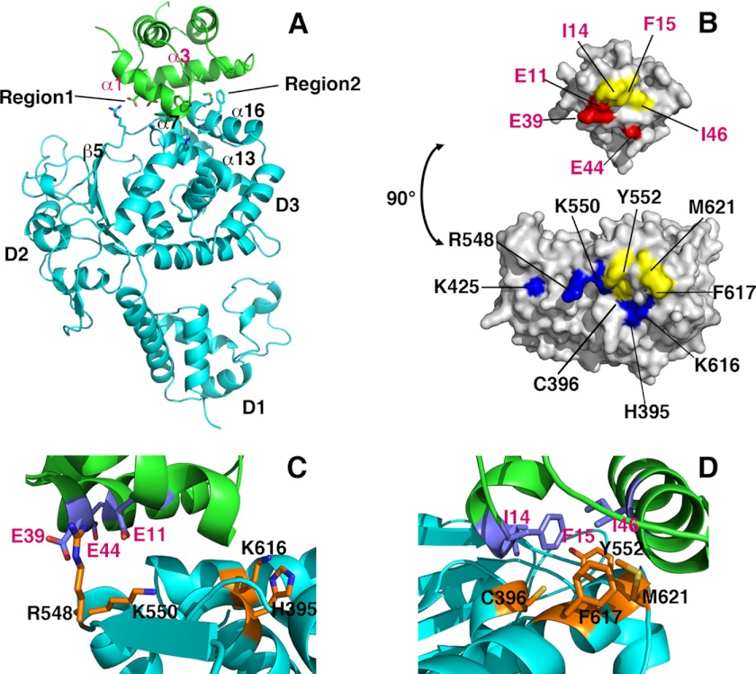
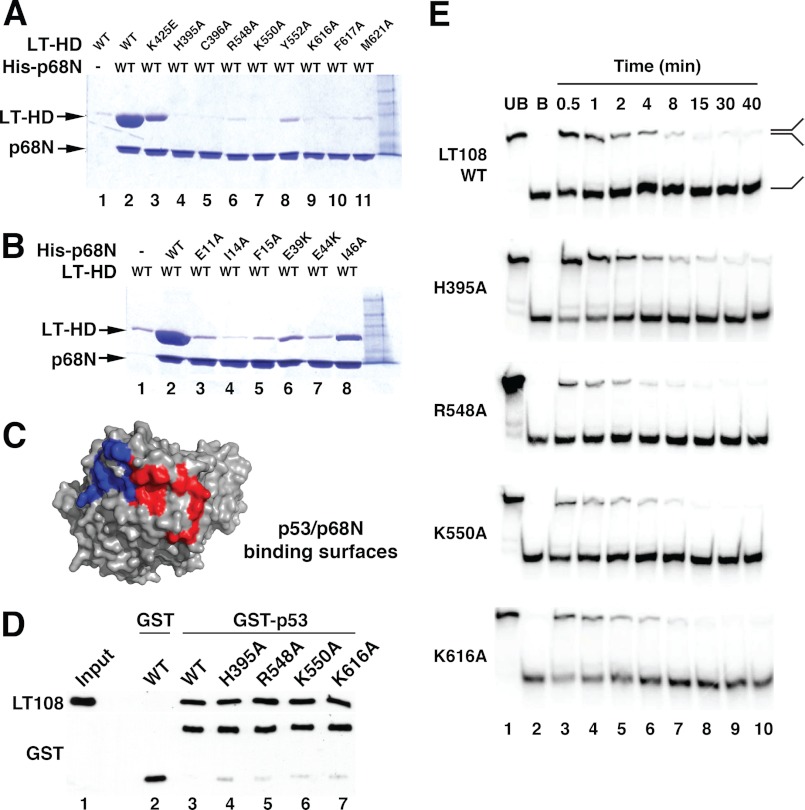
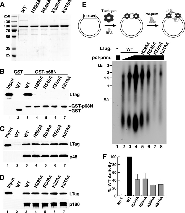
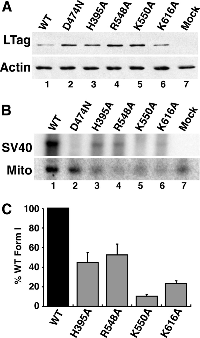
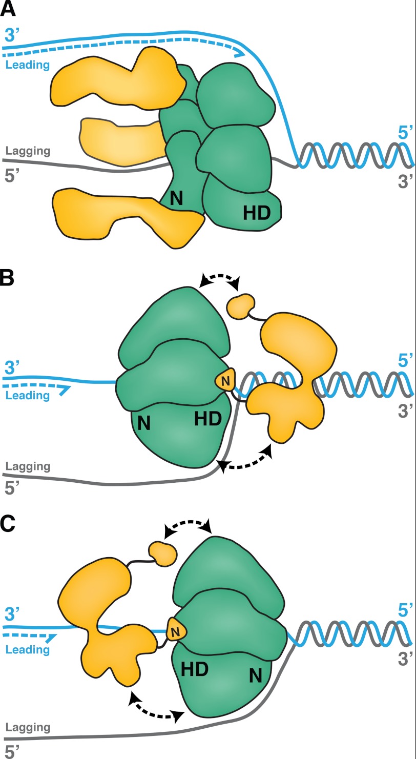
DNA polymerase α-primase (Pol-prim) plays an essential role in eukaryotic DNA replication, initiating synthesis of the leading strand and of each Okazaki fragment on the lagging strand. Pol-prim is composed of a primase heterodimer that synthesizes an RNA primer, a DNA polymerase subunit that extends the primer, and a regulatory B-subunit (p68) without apparent enzymatic activity. Pol-prim is thought to interact with eukaryotic replicative helicases, forming a dynamic multiprotein assembly that displays primosome activity. At least three subunits of Pol-prim interact physically with the hexameric replicative helicase SV40 large T antigen, constituting a simple primosome that is active in vitro. However, structural understanding of these interactions and their role in viral chromatin replication in vivo remains incomplete. Here, we report the detailed large T antigen-p68 interface, as revealed in a co-crystal structure and validated by site-directed mutagenesis, and we demonstrate its functional importance in activating the SV40 primosome in cell-free reactions with purified Pol-prim, as well as in monkey cells in vivo.


Figures

FIGURE 1.
FIGURE 1.
Schematic diagram of the interactions of hexameric LTag with Pol-prim. A, modular architecture of SV40 LTag (27, 51). Each of the three structured domains is sufficient for its biochemical activity as follows: DnaJ chaperone domain (J), origin DNA binding domain (OBD), and helicase domain (HD), composed of the zinc (Zn) and ATPase subdomains, are boxed. Flexible linker regions are indicated as lines. B, hexameric helicase domain of LTag (light gray) contacts Pol-prim (dark gray) through at least three subunits (curved arrows). The N-terminal regions (N) of p180 and p68 are dispensable for enzymatic activity but interact physically with LTag (9, 17, 23, 60).
FIGURE 2.
FIGURE 2.
Overall structure of the LTag-p68N complex. The C-terminal (A) and side (B) views of the LTag helicase domain hexamer in complex with p68N are shown. The six molecules of p68N are colored in green, and each LTag subunit is in a discrete color. For comparison, the C-terminal (C) and side (D) views of LTag hexamer alone are shown.
FIGURE 3.
FIGURE 3.
Detailed LTag-p68N interface interactions. A, ribbon illustration of the complex structure of one LTag molecule (in cyan) binding to one p68N (in green). LTag domains D1, D2, and D3 are indicated. Secondary structures involved in the interaction of both proteins are labeled for LTag and p68N, respectively. Two regions featuring hydrophobic and electrostatic interactions are indicated. B, surface representation of LTag (bottom) and p68N (top), showing the interface areas on both proteins. The residues involved in the interface contacts are colored as follows: hydrophobic residues in yellow, positively charged residues in blue, and negatively charged residues in red. The LTag Lys-425 is not part of the p68N-binding residues, but it is located immediately next to the interface. C and D, close-up views of the detailed LTag-p68N interactions within region 1 (C) and region 2 (D), showing the charge-charge interactions in region 1 and the hydrophobic interactions in region 2, respectively.
FIGURE 4.
FIGURE 4.
Mutational analysis of the LTag-p68N interface and functional validation. A, pulldown assays were performed to evaluate the effect of LTag mutations of the residues within the interface on binding to p68N. Lane 1, LTag helicase domain (LT-HD) retained on Ni resin in the absence of His6-p68N; lane 2, the LTag retained on His6-p68N-bound Ni resin; lanes 3–11, mutant LTags (as marked) retained on His6-p68N-bound Ni resin. B, pulldown assays were performed to evaluate the effect of p68N mutations of the residues within the interface on binding of LTag. Lane 1, LTag retained on Ni resin in the absence of His6-p68N; lane 2, LTag retained on His6-p68N-bound Ni resin; lanes 3–8, LTags retained on Ni resin bound to mutant His6-p68Ns (as marked). For the pulldown assays in A and B, the input LTag for initial incubation for each lane was 100 μg (see “Experimental Procedures”). C, binding interfaces on LTag for p53 (red) (39) and for p68 (blue) (Fig. 3) are adjacent but distinct. D, glutathione-agarose beads bound to either GST (lane 2) or GST-p53 DBD (lanes 3–7) were incubated with LT108 WT or substitution proteins as indicated. Retained proteins were visualized by Western blot with the indicated antibodies. Lane 1, 15% of the LT108 input amounts used in lanes 2–7. E, helicase activities of LT108 mutants that have disrupted p68N binding were assayed over a time course of 30 s to 40 min. Lanes 1 and 2 contain unboiled (UB) and boiled (B) DNA substrate.
FIGURE 5.
FIGURE 5.
Specific role of LTag-p68 interaction in primosome activity. A, purified full-length WT and the indicated mutant LTags were separated by SDS-PAGE and stained with Coomassie Brilliant Blue. Marker proteins are shown at left. B, glutathione-agarose beads bound to either GST (lane 2) or GST-p68N (lanes 3–7) were incubated with full-length WT or the indicated point mutant LTags. Retained proteins were analyzed by Western blot with the indicated antibodies. Lane 1, 15% of the LTag input used for pulldowns in lanes 2–7. C and D, FLAG beads without (lane 2) or with bound p48/His6-FLAG×2-p58 heterodimer (C) or SJK237-31-Sepharose beads without (lane 2) or with bound p180 (D) were incubated with soluble, purified full-length WT or the point mutant LTags as indicated. Lane 1 shows 7.5% of the LTag input used for pulldowns in lanes 2–7. E, initiation of SV40 DNA replication initiation was assayed in monopolymerase reactions containing purified LTag, RPA, topoisomerase, and Pol-prim (diagram). Radiolabeled products of reactions lacking LTag (lane 1) or containing 300 ng of WT or the indicated mutant LTags (lanes 2–8) and varying amounts of Pol-prim (lane 2, 125 ng; lane 3, 250 ng; lanes 1 and 4–8, 500 ng) were analyzed by denaturing gel electrophoresis and phosphorimaging. DNA size markers are indicated (kb). F, initiation activity from three independent experiments as in E was quantified by phosphorimaging, and the activity of each mutant LTag was expressed relative to that of the WT LTag activity in each experiment. No T, as a negative control, LTag was omitted from the sample. Error bars indicate standard deviation.
FIGURE 6.
FIGURE 6.
LTag-p68 interaction is required to replicate SV40 chromatin in monkey kidney cells. A, whole cell extracts were prepared from BSC40 cells transfected 24 h earlier with genomic SV40 DNA encoding WT LTag, the Walker B mutant LTag D474N, or the indicated mutant LTags with defects in p68 binding. A 4-μg sample of total protein from each extract was analyzed by SDS-PAGE and Western blotting to evaluate LTag expression. Actin served as control for equal protein loading. B, low molecular weight DNAs were extracted from a parallel experiment (A) at 48 h after transfection and analyzed by Southern blotting with a radiolabeled SV40 DNA probe and with a human mitochondrial (Mito) DNA probe as a recovery control. C, radiolabeled signals for SV40 form I DNA replication product and mitochondrial DNA in each lane were quantified using phosphorimaging. Replication signals were corrected for sample to sample recovery variations using mitochondrial DNA and for background using the helicase-dead D474N mutant and were compared with that of WT in the same experiment, as detailed under “Experimental Procedures.” The graph shows the average value from two independent transfection experiments. Error bars represent standard deviation.
FIGURE 7.
FIGURE 7.
Variations on primosome architecture. A, in a model prokaryotic primosome, the E. coli replicative helicase, DnaB (green), translocates along the lagging strand template with 5′ to 3′ polarity, its C-terminal helicase domains (HD) facing toward the duplex DNA (gray/cyan). Three molecules of primase, DnaG (yellow), interact with the N-terminal (N) domain of DnaB and trail behind. B, LTag hexamer (green) interacts through its C-terminal helicase domains (HD) with at least three subunits of Pol-prim (yellow). In one possible model of the SV40 primosome, the entire central channel of the hexameric helicase encircles one ssDNA, with the helicase domains facing toward the duplex as LTag translocates with 3′ to 5′ polarity. We suggest that Pol-prim docking on LTag (black double arrows) positions primase to bind the lagging strand template. C, in another possible model, the N-terminal domains of LTag could face the DNA duplex.

Similar articles

Cited by

References

    1. Hamdan S. M., Richardson C. C. (2009) Motors, switches, and contacts in the replisome. Annu. Rev. Biochem. 78, 205–243 - PubMed
    1. Hamdan S. M., van Oijen A. M. (2010) Timing, coordination, and rhythm. Acrobatics at the DNA replication fork. J. Biol. Chem. 285, 18979–18983 - PMC - PubMed
    1. Langston L. D., Indiani C., O'Donnell M. (2009) Whither the replisome. Emerging perspectives on the dynamic nature of the DNA replication machinery. Cell Cycle 8, 2686–2691 - PMC - PubMed
    1. Patel S. S., Pandey M., Nandakumar D. (2011) Dynamic coupling between the motors of DNA replication. Hexameric helicase, DNA polymerase, and primase. Curr. Opin. Chem. Biol. 15, 595–605 - PMC - PubMed
    1. Costa A., Ilves I., Tamberg N., Petojevic T., Nogales E., Botchan M. R., Berger J. M. (2011) The structural basis for MCM2–7 helicase activation by GINS and Cdc45. Nat. Struct. Mol. Biol. 18, 471–477 - PMC - PubMed

Publication types