Review
. 2012 Sep;53(9):1800-10.
doi: 10.1194/jlr.R028290. Epub 2012 Jun 14.

Lipid droplet formation on opposing sides of the endoplasmic reticulum

Affiliations
Review

Lipid droplet formation on opposing sides of the endoplasmic reticulum

Stephen L Sturley et al. J Lipid Res. 2012 Sep.

Abstract

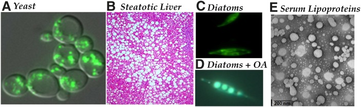
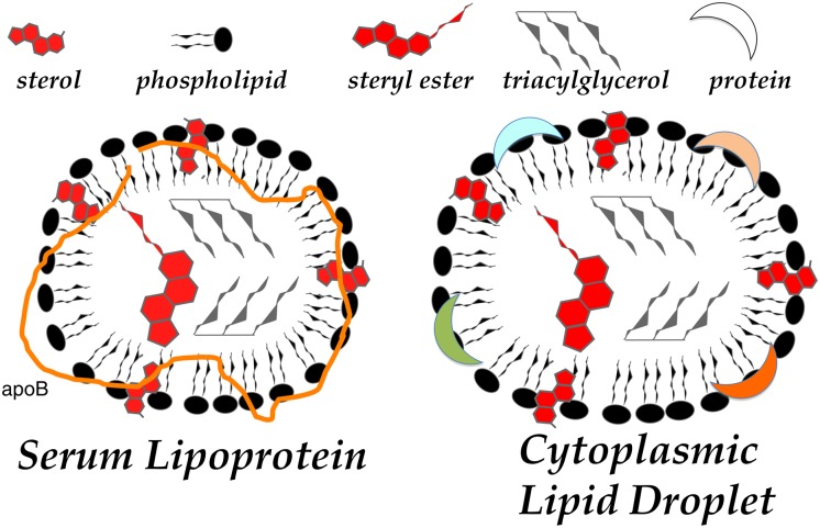
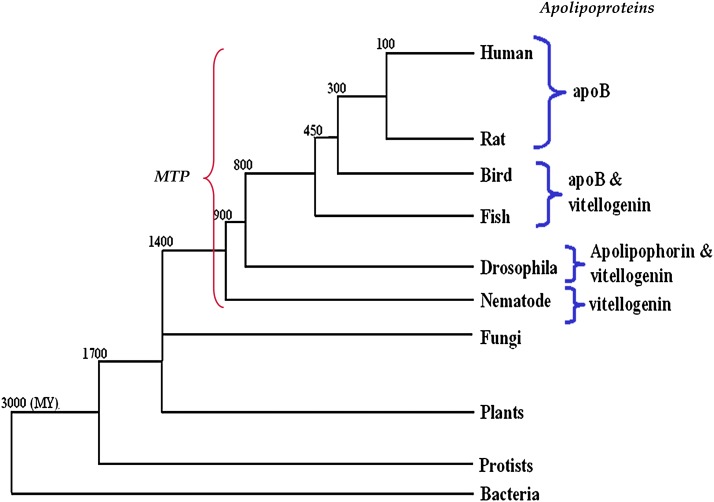
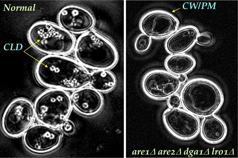
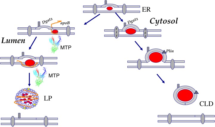
In animal cells, the primary repositories of esterified fatty acids and alcohols (neutral lipids) are lipid droplets that form on the lumenal and/or cytoplasmic side of the endoplasmic reticulum (ER) membrane. A monolayer of amphipathic lipids, intermeshed with key proteins, serves to solubilize neutral lipids as they are synthesized and desorbed. In specialized cells, mobilization of the lipid cargo for delivery to other tissues occurs by secretion of lipoproteins into the plasma compartment. Serum lipoprotein assembly requires an obligate structural protein anchor (apolipoprotein B) and a dedicated chaperone, microsomal triglyceride transfer protein. By contrast, lipid droplets that form on the cytoplasmic face of the ER lack an obligate protein scaffold or any required chaperone/lipid transfer protein. Mobilization of neutral lipids from the cytosol requires regulated hydrolysis followed by transfer of the products to different organelles or export from cells. Several proteins play a key role in controlling droplet number, stability, and catabolism; however, it is our premise that their formation initiates spontaneously, solely as a consequence of neutral lipid synthesis. This default pathway directs droplets into the cytoplasm where they accumulate in many lipid disorders.


Figures

Fig.1.
Fig.1.
Lipid droplets are ubiquitous. All eukaryotes produce and often accumulate neutral lipids as cytoplasmic or secreted particles. A: In budding yeast (Saccharomyces cerevisiae), lipid droplets (envisaged here by fluorescence staining with Nile red) accumulate as cells approach stationary phase. B: The accumulation of fatty deposits in the liver (oil red O staining; Liu and Sturley, unpublished data) is the pathological determinant of steatosis and nonalcoholic fatty liver disease (NAFLD). C, D: Lipid deposition in algae (e.g., diatoms stained with Nile red grown in media lacking and containing free fatty acids, respectively; Ruggles and Sturley, unpublished data) form the basis of our oil reserve. E: A similar structure, triglyceride-rich lipoproteins (negative staining, electron micrograph; Iqbal and Hussain, unpublished data), such as very low density lipoproteins, form in the ER lumen and accumulate in plasma of many patients at risk for coronary artery disease. OA, oleic acid.
Fig.2.
Fig.2.
Cytoplasmic lipid droplet and serum lipoprotein structures. A commonality to cytoplasmic and serum lipid carriers that reflects their shared origin is the membrane of the endoplasmic reticulum. Both particles bud from the ER membranes as neutral lipid is synthesized and approaches insolubility in the membrane. Insolubility is overcome by micellar association with phospholipids and, to a lesser extent, sterols, from the ER and proteins, such as apoB (shown), acyltransferases, lipases, or perilipins (generically indicated by crescents).
Fig.3.
Fig.3.
Gene families involved in terminal steps of neutral lipid biosynthesis. A: Acyl-CoA-dependent acyltransferases. Two unrelated gene families (ACAT and DGAT2) encode acyltransferases with similar activities that transfer the acyl moiety of acyl-CoA. B: Acyl-CoA-independent acyltransferases. In yeast and likely mammalian cells, triglyceride biosynthesis proceeds in the absence of members of the ACAT and DGAT2 gene families. Members of the PDAT gene family perform this reaction independently of acyl-CoA by using phospholipids, such as phosphatidylcholine, as acyl donors. FA-CoA, fatty acyl CoEnzyme A; SE, steryl ester; CoA, CoEnzyme A; DAG, diacylglycerol; TAG, triacylglycerol; LCA, long chain alcohol; WE, wax ester; LCAA, long chain dialcohol; WDE, wax diester, PL, phospholipid. Gene symbols represent establish nomenclature for human acyltransferases and yeast orthologs (in parentheses).
Fig.4.
Fig.4.
A: Deposition of neutral lipids into the cytosol. In all eukaryotic cells and some bacteria, neutral lipids are deposited into the cytoplasm as phopholipid monolayers surrounding an insoluble core of oil. In mammalian cells and most other eukaryotes, the only necessary factor appears to be neutral lipid biosynthetic reactions, such as the DGAT and PDAT reactions. B: Deposition of neutral lipids into the vesicular secretion pathway. In certain specialized tissues and cells (e.g., hepatocytes, enterocytes), neutral lipids are actively packaged into lipoprotein particles by chaperones and are ultimately secreted. Acyltransferases (Pdat, Acat1, Acat2, Dgat1, Dgat2), lumenal transfer/anchor proteins (MTP and apoB), and neutral lipids (SE and TG) are as described in the text. In some instances, the lipid core of the lipid particle is heterogeneous and of obscure origin in terms of acyltransferase isoform (which is depicted without a numerical assignment).
Fig.5.
Fig.5.
Evolution of large lipid transfer proteins. Members of large lipid transfer proteins, such as MTP, apoB, and vitellogenin, share sequence homology. Sequence relationships depict a coevolution that correlates to the appearance of circulatory systems in insects and nematodes. Note that MTP is present in all organisms and is perhaps the first protein to evolve. Throughout evolution, different lipid carrier proteins have arisen, including vitellogenin, apolipophorin, and apoB. Approximate time of divergence is shown at break points. Modified from Ref. .
Fig.6.
Fig.6.
Polarizing light microscopy of yeast strains. Budding yeast imaged with the liquid crystal polarizing microscope (LC-PolScope, Caliper Life Sciences). Cells are surrounded by a highly birefringent cell wall, shown in white in these retardance images. Inside cells, the highly birefringent shell of the lipid droplets (blue arrows) are also visible. In acyltransferase (neutral lipid-deficient, mutant cells shown on the right), lipid droplets are absent. Unlike traditional polarized light microscopes, the so-called retardance image generated by the LC-PolScope represents birefringent structures in shades of gray that are proportional to the amount of birefringence, irrespective of the orientation of the structure. The technical for the amount of birefringence measured by the LC-PolScope is retardance and is expressed as a distance. In these images, white corresponds to 3 nm retardance, medium gray is 1.5 nm retardance. The more that highly ordered lipids and embedded membrane proteins surround the lipid droplets, the more retardance is measured. Images provided by Rudolf Oldenbourg, Marine Biology Laboratory, Woods Hole, MA.
Fig.7.
Fig.7.
Models for cytoplasmic droplet and serum lipoprotein formation in specialized cells. Lipid droplets form on either side of the ER membrane. Import into the cytoplasm reflects the thermodynamic nature of neutral lipids that oil-out the ER membrane. It is unclear whether the droplets are tethered to the ER or migrate away (as shown here). Accessory proteins, such as the fat storage-inducing transmembrane proteins (FIT) and BSCL2/Seipin (SEI), then stabilize and regulate the nascent particles. Dgat2, as a consequence of putative CLD localization, may initiate or supplement core neutral lipid loading while Perilipins (Plin) regulate hydrolysis by specific lipases. In the liver, neutral lipid deposition also occurs in the lumen of the ER and requires a defined set of proteins, specifically an apoB scaffold that acquires lipids, such as triglyceride, as both molecules are produced. The lipid transfer protein MTP, a complex of a transfer polypeptide and the molecular chaperone protein disulphide isomerase, is essential for efficient mobilization of these lipids.

References

    1. Wilson E. B. 1896. The Cell in Development and Inheritance Macmillan, New York.
    1. Chao F. F., Stiers D. L., Ontko J. A. 1986. Hepatocellular triglyceride synthesis and transfer to lipid droplets and nascent very low density lipoproteins. J. Lipid Res. 27: 1174–1181. - PubMed
    1. Turkish A., Sturley S. L. 2007. Regulation of triglyceride metabolism. I. Eukaryotic neutral lipid synthesis: “Many ways to skin ACAT or a DGAT”. Am. J. Physiol. Gastrointest. Liver Physiol. 292: G953–G957. - PubMed
    1. Stone S. J., Myers H. M., Watkins S. M., Brown B. E., Feingold K. R., Elias P. M., Farese R. V., Jr 2003. Lipopenia and skin barrier abnormalities in DGAT2-deficient mice. J. Biol. Chem. 279:11767–11176.
    1. Harris C. A., Haas J. T., Streeper R. S., Stone S. J., Kumari M., Yang K., Han X., Brownell N., Gross R. W., Zechner R., et al. 2011. DGAT enzymes are required for triacylglycerol synthesis and lipid droplets in adipocytes. J. Lipid Res. 52: 657–667. - PMC - PubMed

Publication types

LinkOut - more resources