Review
. 2012 Jun;191(2):347-87.
doi: 10.1534/genetics.111.132886.

Cell polarization and cytokinesis in budding yeast

Affiliations
Review

Cell polarization and cytokinesis in budding yeast

Erfei Bi et al. Genetics. 2012 Jun.

Abstract

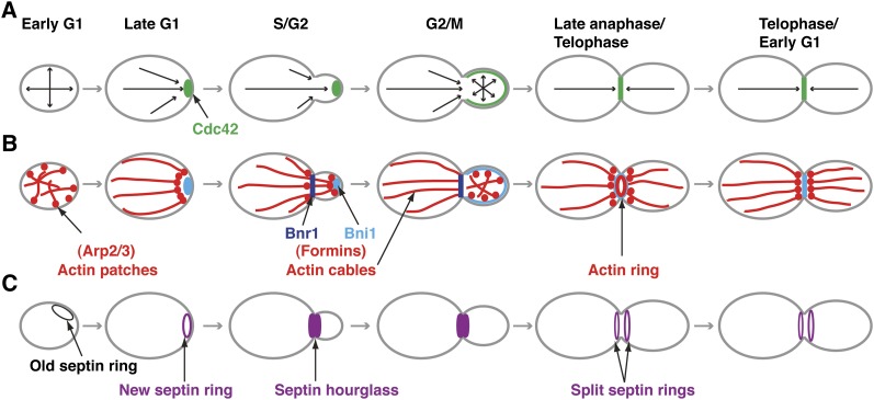
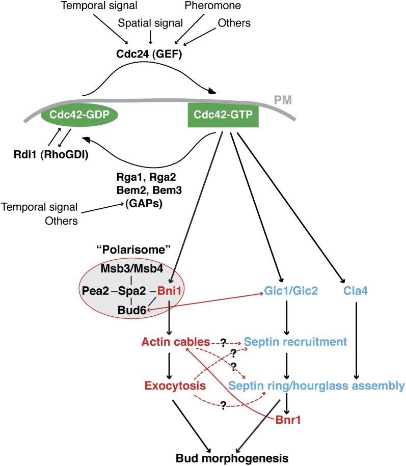
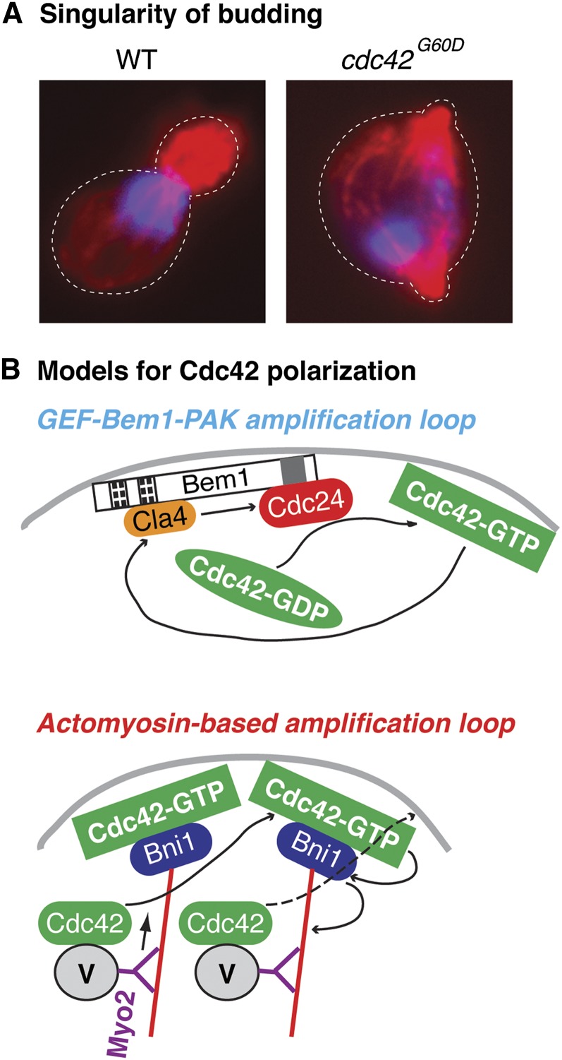
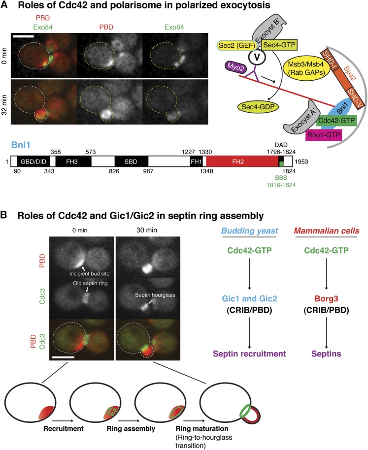
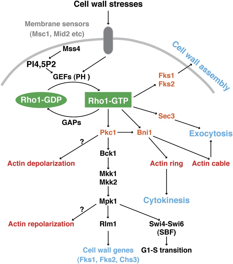
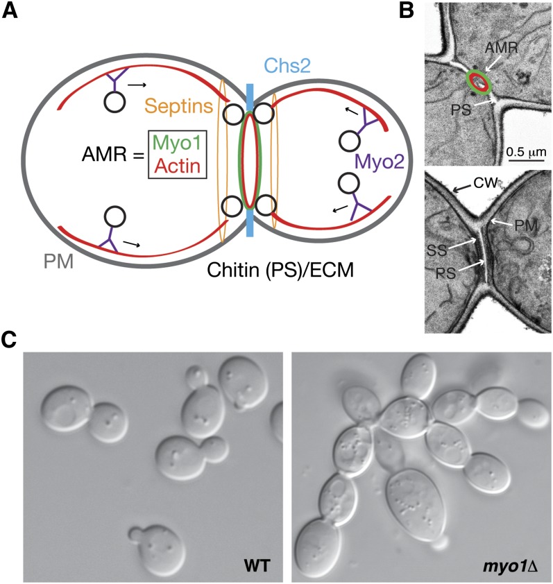
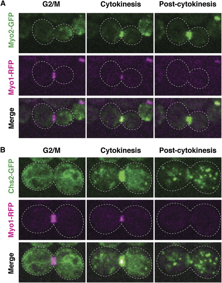
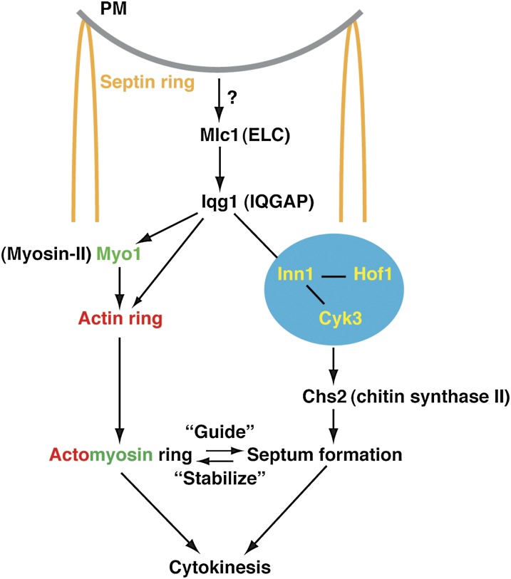
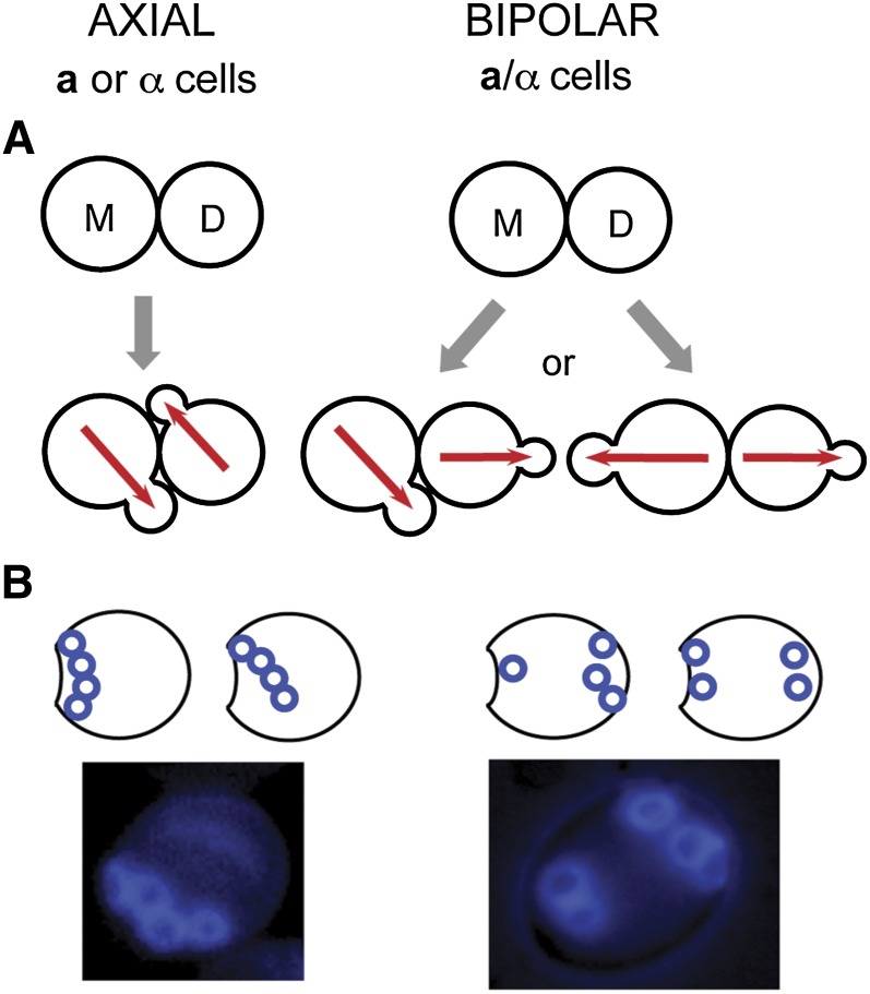
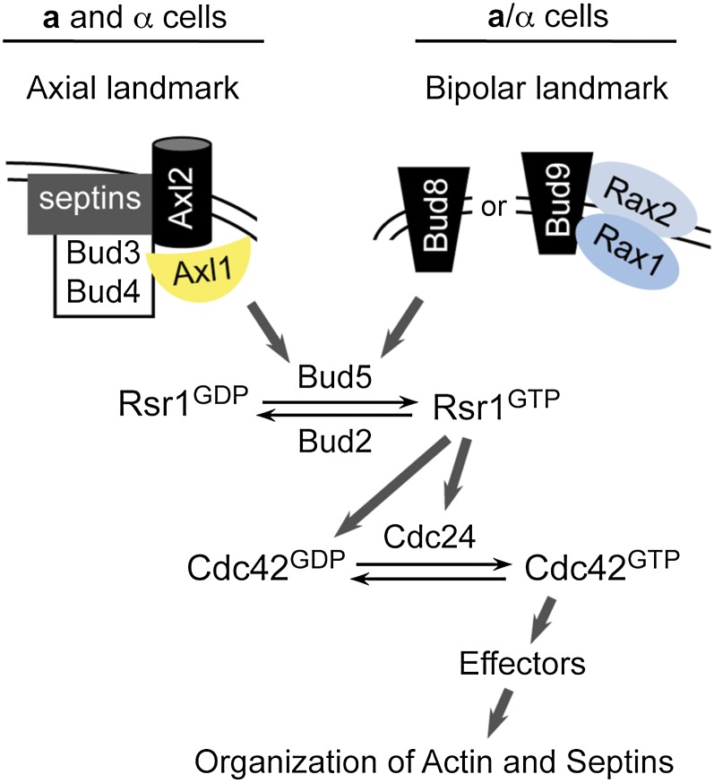
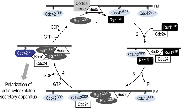
Asymmetric cell division, which includes cell polarization and cytokinesis, is essential for generating cell diversity during development. The budding yeast Saccharomyces cerevisiae reproduces by asymmetric cell division, and has thus served as an attractive model for unraveling the general principles of eukaryotic cell polarization and cytokinesis. Polarity development requires G-protein signaling, cytoskeletal polarization, and exocytosis, whereas cytokinesis requires concerted actions of a contractile actomyosin ring and targeted membrane deposition. In this chapter, we discuss the mechanics and spatial control of polarity development and cytokinesis, emphasizing the key concepts, mechanisms, and emerging questions in the field.


Figures

Figure 1
Figure 1
Cdc42, cell growth, and cytoskeletal polarizations during the cell cycle. (A) Cdc42 (green) localization and the direction of cell growth (arrows) are indicated. (B) Actin organization during the cell cycle. Branched actin filaments in actin patches, nucleated by the Arp2/3 complex, regulate endocytosis. Linear actin cables, nucleated by the formins Bni1 and Bnr1, guide polarized exocytosis. The actin ring, nucleated by the formins (mainly Bni1), is involved in cytokinesis. (C) Septin organization during the cell cycle. Polarized Cdc42 directs septin recruitment to the incipient bud site to form a cortical ring. Upon bud emergence, the septin ring is expanded into an hourglass spanning the entire mother-bud neck. At the onset of cytokinesis, the MEN triggers the splitting of the hourglass into two cortical rings. Modified from (Park and Bi 2007) with permission.
Figure 2
Figure 2
Regulation of Cdc42 and a model for Cdc42-controlled actin cable and septin ring assembly. Internal or external cues activate the GEF Cdc24, which converts Cdc42 to its active GTP-bound state. Activated Cdc42 binds to its effectors to promote actin cable and septin ring assembly (see text for details). Cdc42-GAPs (Rga1, Rga2, Bem2, and Bem3), which are also regulated by internal and external cues, inactivate Cdc42 by stimulating its intrinsic GTPase activity. Rdi1 (Rho GDI) cycles Cdc42-GDP between the PM and the cytosol. Modified from (Park and Bi 2007) with permission.
Figure 3
Figure 3
Singularity of polarization. (A) During each cell cycle, wild-type (WT) cells undergo a single round of budding. (Blue, DNA; red, actin.) In contrast, cells with the hyperactive cdc42G60D allele can bud more than once per cell cycle. (B) Models for Cdc42 polarization. (Top) The scaffold protein Bem1 binds Cdc24 and Cla4 through distinct domains. Cdc24 increases Cdc42-GTP concentration at the polarization site. Increased Cdc42-GTP binds Cla4, which phosphorylates and may activate Cdc24, creating a positive feedback loop. (Bottom) Cdc42-GTP binds the formin Bni1, causing polarization of actin cables toward the growth site. The cables mediate myosin-V (Myo2)-dependent transport of vesicles (V) carrying Cdc42 as a “cargo.” The released Cdc42 will be converted to its active form, which binds to Bni1, leading to more actin cables and more Cdc42 transport, generating a positive feedback loop.
Figure 4
Figure 4
Roles of Cdc42 in polarized exocytosis and septin ring assembly. (A) Roles of Cdc42 and polarisome in polarized exocytosis. (Left) Spatial relationship between Cdc42-GTP and exocytosis. Gic2-PBD (p21-binding domain) fused to tdTomato is a reporter for Cdc42-GTP (PBD) and Exo84-GFP is a marker for polarized exocytosis. Localizations from a time-lapse series are shown. (Right) Spa2, the scaffold protein of the polarisome, binds Bni1 and the Rab GAPs Msb3/Msb4 through distinct domains. Cdc42 and Rho1 control the localization of the exocyst by interacting with the exocyst components Sec3 and Exo70 (Exocyst A′). Cdc42 and Rho1 also control the localization and activation of Bni1, which promotes actin cable assembly toward the site of cell growth. Vesicles (V) carrying the Rab Sec4, Sec2 (the GEF for Sec4), and part of the exocyst (Exocyst B′, which consists of Sec5, Sec6, Sec8, Sec10, Sec15, and Exo70 and Exo84) are transported by myosin-V (Myo2) along the actin cables. Upon vesicle arrival, intact exocyst is assembled by the interaction of exocyst A′ with exocyst B′, which then promotes vesicle tethering to the PM and subsequent membrane fusion. GTP hydrolysis of Sec4 is stimulated by Msb3 and Msb4. (Bottom) Domains of Bni1 (Mosley and Goode 2005, 2006; Ozaki-Kuroda et al. 2001). GBD/DID, GTPase-binding domain/diaphanous inhibitory domain; FH 1-3, formin homology 1-3; SBD, Spa2-binding domain; DAD, diaphanous auto-regulatory domain; BBS, Bud6-binding site. (B) Roles of Cdc42 in septin ring assembly. (Left) Spatial relationship between Cdc42-GTP (PBD) and the septins at the incipient bud site (0 min) and after bud emergence (30 min). (Bottom) Stages of septin ring assembly. (Right) Comparison of yeast and mammalian pathways.
Figure 5
Figure 5
Rho1 in cell wall remodeling, exocytosis, and cytokinesis. Cell wall stresses are sensed by membrane proteins (gray), which activate Rho1-GEFs. Mss4 synthesizes PI(4,5)P2 at the PM (gray line), which recruits GEFs via their PH domains. Activated Rho1 controls cell wall assembly by directly activating glucan synthases (Fks1 and Fks2) and by activating the Pkc1-MAPK–mediated CWI pathway, which induces expression of cell-wall synthetic enzymes. Rho1 also regulates exocytosis and cytokinesis via Sec3 and Bni1. In response to heat stress, Rho1 and Pkc1 cause transient actin depolarization, and the MAPK pathway promotes subsequent actin repolarization. Modified from (Park and Bi 2007) with permission.
Figure 6
Figure 6
Events of cytokinesis. (A) Coupling of AMR contraction to membrane trafficking and primary septum (PS) formation during cytokinesis. Myo1, myosin-II; Myo2, myosin-V; Chs2, a transmembrane chitin synthase responsible for PS formation. Black circles, post-Golgi vesicles; red lines, actin cables. (B) EM visualization of the primary and secondary septa. SS, secondary septa; CW, cell wall. An AMR is drawn to illustrate its spatial relationship with the PS during cytokinesis. (C) Cytokinesis defects of myo1Δ cells. Unlike wild-type (WT) cells, which are either unbudded or single-budded, myo1Δ cells form extensive chains or clusters, indicative of cytokinesis and cell-separation defects. Micrographs in B and for WT cells in C were published previously (Fang et al. 2010).
Figure 7
Figure 7
Spatiotemporal coupling of actomyosin ring contraction with membrane trafficking and septum formation during cytokinesis. (A) Spatiotemporal relationship between Myo1 (magenta) and Myo2 (green) before, during, and after cytokinesis. (B) Spatiotemporal relationship between Myo1 (magenta) and Chs2 (green) before, during, and after cytokinesis.
Figure 8
Figure 8
A molecular model for cytokinesis in budding yeast. At the onset of cytokinesis, the septin hourglass is split into two cortical rings (light brown), Mlc1 and Iqg1 maintain Myo1 at the division site, and all three proteins are required for AMR assembly and constriction. Iqg1 is also involved in septum formation, possibly by interacting with Inn1. Inn1 interacts with Hof1 and Cyk3 to somehow “activate” Chs2 for PS formation. AMR “guides” membrane deposition and septum formation whereas the latter “stabilizes” the AMR and its constriction. Efficient cytokinesis requires spatiotemporal coordination of the AMR and septum formation.
Figure 9
Figure 9
Patterns of bud-site selection in S. cerevisiae. (A) Axial and bipolar patterns of cell division. Red arrows denote polarization axes. (B) The patterns of bud scars on the yeast cell surface resulting from the two different modes of budding. On each cell a single birth scar marks where the cell detached from its mother (M). A bud scar shown as a blue ring marks a division site on the mother cell surface. Bud scars can be visualized by staining with the dye Calcofluor (as shown) or by scanning electron microscopy. In the axial pattern, scars form a continuous chain. In the bipolar pattern, scars cluster around the birth pole (proximal pole) and the pole opposite the birth end (distal pole). Modified from (Park and Bi 2007) with permission.
Figure 10
Figure 10
Pathways governing axial and bipolar budding in haploid (a or α) and diploid (a/α) cells. Although physical interaction has been demonstrated in some cases, many interactions are postulated on the basis of genetic and localization data. All proteins do not necessarily interact at the same time. See text for further details. Modified from (Park and Bi 2007) with permission.
Figure 11
Figure 11
A model for how the Rsr1 GTPase cycle directs polarity establishment to a specific site. (Step 1) Bud5, which is recruited to a specific site by a spatial landmark, catalyzes GDP/GTP exchange on Rsr1. (Step 2) Rsr1-GTP associates with Cdc24 and Cdc42 and guides them to the presumptive bud site. (Step 3) Bud2 stimulates Rsr1 to hydrolyze its bound GTP. (Step 4) Cdc24 no longer interacts with Rsr1 and catalyzes the exchange of GDP for GTP on Cdc42, leading to local Cdc42 activation. Cdc42-GTP then triggers actin assembly and exocyst localization to establish an axis of cell polarization. Bud5 at the presumptive bud site may convert Rsr1 to a GTP-bound state (dashed line), allowing for another GTPase cycle. Homotypic Rsr1–Rsr1 interaction and heterotypic Rsr1–Cdc42 interaction may stabilize these GTPases at a single site, contributing to proper bud-site selection and polarity establishment. Modified from (Park and Bi 2007) with permission.

References

    1. Abe M., Qadota H., Hirata A., Ohya Y., 2003. Lack of GTP-bound Rho1p in secretory vesicles of Saccharomyces cerevisiae. J. Cell Biol. 162: 85–97 - PMC - PubMed
    1. Adames N., Blundell K., Ashby M. N., Boone C., 1995. Role of yeast insulin-degrading enzyme homologs in propheromone processing and bud site selection. Science 270: 464–467 - PubMed
    1. Adamo J. E., Rossi G., Brennwald P., 1999. The Rho GTPase Rho3 has a direct role in exocytosis that is distinct from its role in actin polarity. Mol. Biol. Cell 10: 4121–4133 - PMC - PubMed
    1. Adamo J. E., Moskow J. J., Gladfelter A. S., Viterbo D., Lew D. J., et al. , 2001. Yeast Cdc42 functions at a late step in exocytosis, specifically during polarized growth of the emerging bud. J. Cell Biol. 155: 581–592 - PMC - PubMed
    1. Adams A. E., Botstein D., Drubin D. G., 1989. A yeast actin-binding protein is encoded by SAC6, a gene found by suppression of an actin mutation. Science 243: 231–233 - PubMed

Publication types

MeSH terms