

Barrett's Esophagus: Emerging Knowledge and Management Strategies
- PMID: 22701199
- PMCID: PMC3369502
- DOI: 10.1155/2012/814146
Barrett's Esophagus: Emerging Knowledge and Management Strategies
Abstract
The incidence of esophageal adenocarcinoma (EAC) has increased exponentially in the last 3 decades. Barrett's esophagus (BE) is the only known precursor of EAC. Patients with BE have a greater than 40 folds higher risk of EAC compared with the general population. Recent years have witnessed a revolution in the clinical and molecular research related to BE. However, several aspects of this condition remain controversial. Data regarding the true prevalence of BE have varied widely. Recent studies have suggested a lower incidence of EAC in nondysplastic BE (NDBE) than previously reported. There is paucity of prospective data showing a survival benefit of screening or surveillance for BE. Furthermore, the ever-increasing emphasis on healthcare cost containment has called for reexamination of the screening and surveillance strategies for BE. There is a need for identification of reliable clinical predictors or molecular biomarkers to risk-stratify patients who might benefit the most from screening or surveillance for BE. Finally, new therapies have emerged for the management of dysplastic BE. In this paper, we highlight the key areas of controversy and uncertainty surrounding BE. The paper discusses, in detail, the current literature about the molecular pathogenesis, biomarkers, histopathological diagnosis, and management strategies for BE.
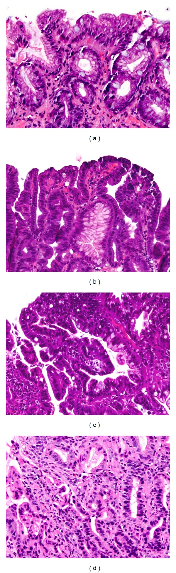
Figures

Similar articles
-
The controversy regarding ablation for Barrett's esophagus without dysplasia.Curr Opin Gastroenterol. 2011 Jul;27(4):368-73. doi: 10.1097/MOG.0b013e328347bafc. Curr Opin Gastroenterol. 2011. PMID: 21577102 Review.
-
Low risk of adenocarcinoma and high-grade dysplasia in patients with non-dysplastic Barrett's esophagus: Results from a cohort from a country with low esophageal adenocarcinoma incidence.United European Gastroenterol J. 2016 Jun;4(3):343-52. doi: 10.1177/2050640615612409. Epub 2015 Oct 30. United European Gastroenterol J. 2016. PMID: 27403300 Free PMC article.
-
Barrett's Registry Collaboration of academic centers in Ireland reveals high progression rate of low-grade dysplasia and low risk from nondysplastic Barrett's esophagus: report of the RIBBON network.Dis Esophagus. 2020 Oct 12;33(10):doaa009. doi: 10.1093/dote/doaa009. Dis Esophagus. 2020. PMID: 32193532
-
Screening and surveillance for Barrett's esophagus: current issues and future directions.Curr Opin Gastroenterol. 2012 Jul;28(4):377-81. doi: 10.1097/MOG.0b013e328353d58e. Curr Opin Gastroenterol. 2012. PMID: 22508325 Free PMC article. Review.
-
Incidence of Progression of Persistent Nondysplastic Barrett's Esophagus to Malignancy.Clin Gastroenterol Hepatol. 2019 Apr;17(5):869-877.e5. doi: 10.1016/j.cgh.2018.08.033. Epub 2018 Sep 11. Clin Gastroenterol Hepatol. 2019. PMID: 30213587
Cited by
-
Wide-area transepithelial sampling in adjunct to forceps biopsy increases the absolute detection rates of Barrett's oesophagus and oesophageal dysplasia: a meta-analysis and systematic review.BMJ Open Gastroenterol. 2020 Sep;7(1):e000494. doi: 10.1136/bmjgast-2020-000494. BMJ Open Gastroenterol. 2020. PMID: 32928869 Free PMC article.
-
Management strategies of Barrett's esophagus.World J Gastroenterol. 2012 Nov 21;18(43):6216-25. doi: 10.3748/wjg.v18.i43.6216. World J Gastroenterol. 2012. PMID: 23180941 Free PMC article. Review.
-
Wide-area transepithelial sampling with computer-assisted 3-dimensional analysis (WATS) markedly improves detection of esophageal dysplasia and Barrett's esophagus: analysis from a prospective multicenter community-based study.Dis Esophagus. 2019 Mar 1;32(3):doy099. doi: 10.1093/dote/doy099. Dis Esophagus. 2019. PMID: 30541019 Free PMC article.
-
Towards screening Barrett's oesophagus: current guidelines, imaging modalities and future developments.Clin J Gastroenterol. 2020 Oct;13(5):635-649. doi: 10.1007/s12328-020-01135-2. Epub 2020 Jun 3. Clin J Gastroenterol. 2020. PMID: 32495144 Free PMC article. Review.
-
Expression of SOX9 and CDX2 in nongoblet columnar-lined esophagus predicts the detection of Barrett's esophagus during follow-up.Mod Pathol. 2015 May;28(5):654-61. doi: 10.1038/modpathol.2014.157. Epub 2014 Nov 21. Mod Pathol. 2015. PMID: 25412842
References
-
- Steevens J, Botterweck AAM, Dirx MJM, Van Den Brandt PA, Schouten LJ. Trends in incidence of oesophageal and stomach cancer subtypes in Europe. European Journal of Gastroenterology and Hepatology. 2010;22(6):669–678. - PubMed
-
- Yousef F, Cardwell C, Cantwell MM, Galway K, Johnston BT, Murray L. The incidence of esophageal cancer and high-grade dysplasia in Barrett’s esophagus: a systematic review and meta-analysis. American Journal of Epidemiology. 2008;168(3):237–249. - PubMed
-
- Pohl H, Welch HG. The role of overdiagnosis and reclassification in the marked increase of esophageal adenocarcinoma incidence. Journal of the National Cancer Institute. 2005;97(2):142–146. - PubMed
-
- Eloubeidi MA, Mason AC, Desmond RA, El-Serag HB. Temporal trends (1973–1997) in survival of patients with esophageal adenocarcinoma in the United States: a glimmer of hope? American Journal of Gastroenterology. 2003;98(7):1627–1633. - PubMed
Grants and funding
LinkOut - more resources
Full Text Sources
Other Literature Sources
