. 2012;7(5):e38005.
doi: 10.1371/journal.pone.0038005. Epub 2012 May 31.

Expression of transient receptor potential ankyrin 1 (TRPA1) and its role in insulin release from rat pancreatic beta cells

Affiliations

Expression of transient receptor potential ankyrin 1 (TRPA1) and its role in insulin release from rat pancreatic beta cells

De-Shou Cao et al. PLoS One. 2012.

Abstract

Objective: Several transient receptor potential (TRP) channels are expressed in pancreatic beta cells and have been proposed to be involved in insulin secretion. However, the endogenous ligands for these channels are far from clear. Here, we demonstrate the expression of the transient receptor potential ankyrin 1 (TRPA1) ion channel in the pancreatic beta cells and its role in insulin release. TRPA1 is an attractive candidate for inducing insulin release because it is calcium permeable and is activated by molecules that are produced during oxidative glycolysis.

Methods: Immunohistochemistry, RT-PCR, and Western blot techniques were used to determine the expression of TRPA1 channel. Ca²⁺ fluorescence imaging and electrophysiology (voltage- and current-clamp) techniques were used to study the channel properties. TRPA1-mediated insulin release was determined using ELISA.

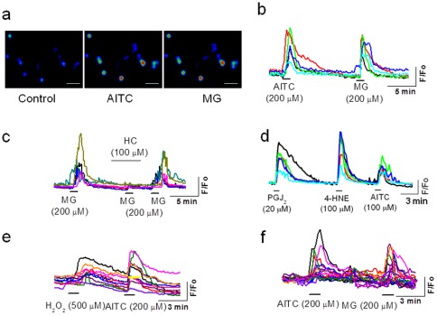
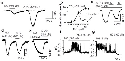
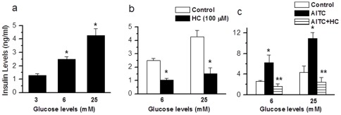
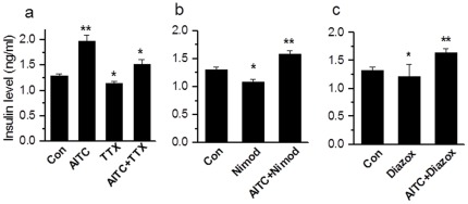
Results: TRPA1 is abundantly expressed in a rat pancreatic beta cell line and freshly isolated rat pancreatic beta cells, but not in pancreatic alpha cells. Activation of TRPA1 by allyl isothiocyanate (AITC), hydrogen peroxide (H₂O₂), 4-hydroxynonenal (4-HNE), and cyclopentenone prostaglandins (PGJ₂) and a novel agonist methylglyoxal (MG) induces membrane current, depolarization, and Ca²⁺ influx leading to generation of action potentials in a pancreatic beta cell line and primary cultured pancreatic beta cells. Activation of TRPA1 by agonists stimulates insulin release in pancreatic beta cells that can be inhibited by TRPA1 antagonists such as HC030031 or AP-18 and by RNA interference. TRPA1-mediated insulin release is also observed in conditions of voltage-gated Na⁺ and Ca²⁺ channel blockade as well as ATP sensitive potassium (K(ATP)) channel activation.

Conclusions: We propose that endogenous and exogenous ligands of TRPA1 cause Ca²⁺ influx and induce basal insulin release and that TRPA1-mediated depolarization acts synergistically with K(ATP) channel blockade to facilitate insulin release.


Conflict of interest statement

Competing Interests: The authors have declared that no competing interests exist.

Figures

Figure 1
Figure 1. Expression of TRPA1 in pancreatic beta cells.
a. RT-PCR shows the expression of TRPA1 mRNA in DRG neurons, whole pancreas (Pan), isolated islets (Isl), a pancreatic beta cell line (RIN), but not in a pancreatic alpha cell line (INR). b. Western blots show the expression of TRPA1 protein in RIN cells and HEK cells heterologously expressing TRPA1. c. Immunostaining of insulin (red), TRPA1 (green), and the co-expression (yellow) in the pancreatic islet (top panel). When the slices were incubated with the TRPA1 antibody after preabsorbing with a peptide used for making the antibody, the TRPA1 staining was considerably reduced (lower panel). The nuclei were stained with DAPI (scale bar=100 µM).
Figure 2
Figure 2. TRPA1-mediated Ca2+
influx in pancreatic beta cells. a, b. Application of AITC and MG induce an increase in intracellular Ca2+ in RIN cells (size of the bar is 100 µM). c. MG-induced Ca2+ influx is inhibited by TRPA1 antagonist HC030031. d. Ca2+ influx induced by endogenous ligands PGJ2, 4-HNE, and AITC in RIN cells. e. Ca2+ influx induced by H2O2 and AITC in RIN cells. f. AITC-and MG-induced an increase in intracellular Ca2+ in rat cultured primary pancreatic beta cells.
Figure 3
Figure 3. TRPA1-mediated membrane currents in primary pancreatic beta cells.
a. Membrane currents induced by extracellular application of MG and AITC in primary pancreatic beta cells. b. A concentration-response curve of membrane currents induced by MG included in the pipette solution in primary beta cells (EC50=0.59 µM). Lower concentrations (∼1 µM) of MG are sufficient to induce maximal currents when applied intracellulary (inset), but the time to peak with lower concentrations is longer and the desensitization is profound at higher concentrations. c. Currents evoked by intracellular application of MG are reversibly blocked by extracellular application of AP-18. d. Currents elicited by MG and AITC in HEK 293T cells heterologously expressing TRPA1. e. MG-induced currents can be blocked by AP-18. f. Under current clamp conditions, extracellular application of MG depolarizes the membrane and generates action potentials that could be blocked by HC030031. g. Intracellular application of MG causes a robust depolarization and generates action potentials that could be blocked by HC030031.
Figure 4
Figure 4. TRPA1-mediated insulin release in pancreatic beta cell line and primary isolated pancreatic islets.
a,b. Dose-dependent increase in insulin release induced by AITC (0.1–1000 µM, n=7) and MG (0.1–1000 µM, n=5) in RIN cells (* p<0.05). c,d. AITC and MG induce a significant increase (AITC, n=11,* p<0.001; MG, n=10 * p=0.004) in insulin release from primary isolated pancreatic beta cell islets that could be blocked by the specific TRPA1 antagonist AP-18 (AITC+AP-18, n=6, ** p<0.001; MG+AP-18, n=6, ** p=0.008). e. 4-HNE (100 µM)-induced insulin release is inhibited by HC030031 (100 µM) (4-HNE, n=6, * p<0.001; 4-HNE+HC030031, n=3, ** p<0.001). f. PGJ2 (20 µM)-induced insulin release is inhibited by HC030031 (100 µM) (PGJ2, n=6, * p<0.001; PGJ2+HC030031, n=3, ** p<0.001).
Figure 5
Figure 5. Insulin release induced by different concentrations of glucose.
a. Insulin release induced by different concentrations of glucose (6 mM, n=8, * p<0.001; 25 mM, n=9, *p<0.001) b. Insulin release induced by different concentrations of glucose is inhibited by HC030031 (100 µM) (6 mM, n=4, * p<0.001; 25 mM, n=7, * p<0.001, as compared to control). c. Insulin release induced by AITC (200 µM) in different concentrations of glucose is inhibited by HC030031 (100 µM) (6 mM, AITC, n=4, * p<0.01, AITC+HC030031, n=4, ** p<0.001; 25 mM, AITC, n=4, * p=0.023, AITC+HC030031, n=4, ** p<0.01).
Figure 6
Figure 6. TRPA1-mediated insulin release is independent of voltage-gated Na+, Ca2+ and KATP channels.
a. AITC caused a significant increase in insulin release (n=6, ** p<0.01). The basal insulin release is inhibited by incubation of RIN cells with TTX (1 µM) (TTX, n=6, * p<0.05. When challenged with AITC (200 µM), there is a significant increase in insulin release AITC+TTX, n=6, * p<0.05 as compared to TTX. b. In the presence of Ca2+ channel blocker nimodipine (5 µM) basal insulin release is decreased significantly (n=6, * p<0.05), but there is a significant increase when challenged with AITC+nimodipine (n=6,** p<0.01). c. In the presence of KATP channel opener, diazoxide (200 µM), basal insulin release is significantly decreased (n=6, * p<0.05), when challenged with AITC, there is a significant increase in insulin release (n=3, ** p<0.01).
Figure 7
Figure 7. TRPA1-medated insulin release and Ca2+ influx following knockdown of TRPA1 by siRNA
a. RT-PCR after knockdown of TRPA1 by siRNA in control and mock-transfected cells. b. AITC (200 µM)-induced insulin release is significantly reduced after siRNA knockdown of TRPA1 in RIN cells (n=6, * p<0.05). c. AITC (200 µM) and MG (1 mM) induce increase in intracellular Ca2+ levels in mock-transfected RIN cells (upper panel), but not in siRNA treated cells (lower panel). Note that siRNA treated cells responded to KCl (50 mM) similar to that of mock-transfected cells.

References

    1. Nilius B, Owsianik G, Voets T, Peters JA. Transient receptor potential cation channels in disease. Physiol Rev. 2007;87:165–217. - PubMed
    1. Razavi R, Chan Y, Afifiyan, Liu XJ, Wan X, et al. TRPV1+ sensory neurons control beta cell stress and islet inflammation in autoimmune diabetes. Cell. 2006;127:1123–1135. - PubMed
    1. Gram DX, Ahrén B, Nagy, Olsen UB, Brand CL, et al. Capsaicin-sensitive sensory fibers in the islets of Langerhans contribute to defective insulin secretion in Zucker diabetic rat, an animal model for some aspects of human type 2 diabetes. Eur J Neurosci. 2007;25:213–223. - PubMed
    1. Ceppa E, Cattaruzza F, Lyo V, Amadesi S, Pelayo JC, et al. Transient receptor potential ion channels V4 and A1 contribute to pancreatitis pain in mice. Am J Physiol Gastrointest Liver Physiol. 2010;299:G556–571. - PMC - PubMed
    1. Akiba Y, Kato S, Katsube K, Nakamura M, Takeuchi K, et al. Transient receptor potential vanilloid subfamily 1 expressed in pancreatic islet beta cells modulates insulin secretion in rats. Biochem Biophys Res Commun. 2004;321:219–225. - PubMed

Publication types

MeSH terms