. 2012;7(6):e38029.
doi: 10.1371/journal.pone.0038029. Epub 2012 Jun 6.

Active zone protein Bassoon co-localizes with presynaptic calcium channel, modifies channel function, and recovers from aging related loss by exercise

Affiliations

Active zone protein Bassoon co-localizes with presynaptic calcium channel, modifies channel function, and recovers from aging related loss by exercise

Hiroshi Nishimune et al. PLoS One. 2012.

Abstract

The P/Q-type voltage-dependent calcium channels (VDCCs) are essential for synaptic transmission at adult mammalian neuromuscular junctions (NMJs); however, the subsynaptic location of VDCCs relative to active zones in rodent NMJs, and the functional modification of VDCCs by the interaction with active zone protein Bassoon remain unknown. Here, we show that P/Q-type VDCCs distribute in a punctate pattern within the NMJ presynaptic terminals and align in three dimensions with Bassoon. This distribution pattern of P/Q-type VDCCs and Bassoon in NMJs is consistent with our previous study demonstrating the binding of VDCCs and Bassoon. In addition, we now show that the interaction between P/Q-type VDCCs and Bassoon significantly suppressed the inactivation property of P/Q-type VDCCs, suggesting that the Ca(2+) influx may be augmented by Bassoon for efficient synaptic transmission at NMJs. However, presynaptic Bassoon level was significantly attenuated in aged rat NMJs, which suggests an attenuation of VDCC function due to a lack of this interaction between VDCC and Bassoon. Importantly, the decreased Bassoon level in aged NMJs was ameliorated by isometric strength training of muscles for two months. The training increased Bassoon immunoreactivity in NMJs without affecting synapse size. These results demonstrated that the P/Q-type VDCCs preferentially accumulate at NMJ active zones and play essential role in synaptic transmission in conjunction with the active zone protein Bassoon. This molecular mechanism becomes impaired by aging, which suggests altered synaptic function in aged NMJs. However, Bassoon level in aged NMJs can be improved by muscle exercise.


Conflict of interest statement

Competing Interests: The authors have declared that no competing interests exist.

Figures

Figure 1
Figure 1. P/Q-type VDCCs localize at the NMJ active zones.
(A) P/Q-type VDCCs stained with an antibody against the α subunit (Cav2.1, green) in NMJs of sternomastoid muscle from postnatal day 15 wild-type mice stained with Alexa Fluor 594-labeled α-bungarotoxin to label the acetylcholine receptors (AChR, red). The immunoreactivity is absent in the NMJ area of the littermate P/Q-type VDCC knockout mice (Cacna1a−/−) demonstrating the specificity of the immunohistochemical signals. The same result was confirmed in four independent litters. (B) P/Q-type VDCCs were detected inside the primary gutter of endplates labeled with α-bungarotoxin, where motor nerve terminals reside (arrowheads). An YZ-orthogonal view (a single optical plane) of a region indicated by an orange line in A is shown. The nerve is placed toward the top of the Z-axis and the muscle toward the bottom. Some P/Q-type VDCC signals were also detected in the muscle side. (C) P/Q-type VDCCs aligned with the active zone protein Bassoon in NMJs (arrowheads). Sternomastoid muscle of postnatal day 21 wild-type mice was stained using an anti-P/Q-type VDCC α subunit antibody (red), an anti-Bassoon antibody (green), and Alexa Fluor 647-labeled α-bungarotoxin (blue). The maximal projected XY-view of confocal Z-stack is shown in C1. Panels C2 and C3 show YZ- and XZ-orthogonal views (a single optical plane) at the positions indicated by the orange lines in C1. Panels C4 and C5 show a magnified region of C1 indicated by the dotted-box (C4) and orange line inside the dotted box (C5). Many Bassoon and P/Q-type VDCCs signals (white arrowheads) align in the XY-views (C4) and YZ-orthogonal views (C5, single optical plane). In C5, the nerve is placed towards the top, and muscle is placed towards the bottom. (C6) Colocalization analysis of Bassoon and P/Q-type VDCC within NMJ presynaptic terminals of postnatal day 21 wild-type mice by the Manders’ coefficients (M). Some degree of colocalization of these proteins was indicated by the significantly higher Manders’ coefficients M values for Bassoon overlapping with P/Q-type VDCC (0.42±0.04, 3 NMJs, Bsn on PQ) and P/Q-type VDCC overlapping with Bassoon (0.39±0.02, 3 NMJs, PQ on Bsn) compared to the M value for acetylcholine receptor overlapping minimally with neurofilament (0.11±0.03; 6 NMJs, AChR on NF). A significant difference was detected using one-way ANOVA (P = 0.0002). Asterisks indicate significant difference against AChR on NF by Bonferroni post-test. Scale bars: A, 10 µm; B, C, 1 µm.
Figure 2
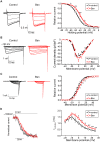
Figure 2. Inactivation properties of the P/Q-type VDCC were suppressed by Bassoon.
(A) Left, inactivation of P/Q-type VDCC (Cav2.1) currents in BHK cells stably expressing VDCCs and transfected with an expression vector pBassoon-IRES2-GFP (Bsn, red) or an empty pIRES2-GFP vector (control, black). The peak amplitudes were normalized for Ba2+ currents elicited by 2-s pulses to 0 mV from a holding potential of –100 mV. Right graph shows inactivation curves for P/Q-type VDCCs with or without Bassoon. The half-inactivation potential was significantly higher in Bassoon expressing cells (–40.0±1.7 mV) compared to controls (–46.1±0.6 mV). (B) I-V relationships of P/Q-type VDCC showed no difference between with or without Bassoon. Left, representative traces are Ba2+ currents of Cav2.1 with or without Bassoon by applying test pluses from –100 mV (holding potential) to –50 mV up to 40 mV in 10 mV increments. Right graph shows current density-voltage (I-V) relationships. (C) The activation property of P/Q-type VDCCs in the presence of Bassoon exhibited a depolarization shift. Left, effects of Bassoon on activation of Cav2.1 currents elicited in BHK cells. Tail currents were elicited by repolarization to –60 mV after 5-ms test pulse from –50 to 55 mV with 5 mV increments. Right, activation curves were determined using these tail currents with or without Bassoon. The half-activation potential was significantly higher in Bassoon expressing cells (–0.7±1.1 mV) compared to controls (–4.0±1.1 mV). (D) Activation kinetics of P/Q-type VDCC currents. Left, tail currents were evoked by 5 ms depolarization from the holding potential (–100 mV) to 0 mV. Right graph shows the activation time constants (τ activation) with or without Bassoon. The activation time constant (τ) increased significantly in the presence of Bassoon at membrane potentials higher than 0 mV. Recordings from eight independent cells were averaged and the mean ± SEM are shown. Asterisks indicate significant difference compared to the controls, *p<0.05, **p<0.01.
Figure 3
Figure 3. Exercise ameliorated the active zone protein Bassoon level in aged NMJs.
(A) NMJs of trained aged rats (trained-aged) showed higher Bassoon signal intensity compared to the NMJs of un-trained aged rats (aged). Two-year-old rats underwent isometric strength training of tongue muscles for two months. Genioglossus muscles of the tongue were sectioned and stained with antibody against Bassoon (active zone marker), neurofilament and SV2 (nerve morphology, NF+SV2), and α-bungarotoxin (acetylcholine receptors, AChR). Highly magnified Bassoon staining of the area indicated by white dotted boxes are shown in the second column from the left (High Mag. Bassoon). NMJs of young rats (young, postnatal day 56) exhibited higher Bassoon signal intensity than aged rats, similar to our previous mouse study . Scale bar: 10 µm. (B) Average signal intensity of Bassoon was significantly higher in NMJs of young rats and trained-aged rats compared to those of untrained-aged rats (mean ± standard error in arbitrary intensity units: young rats, 8.06±1.27; trained-aged rats, 7.81±1.51; and aged rats, 2.51±0.51; four rats in each group). The red bracket indicates a subgroup of NMJs in aged rats with a minuscule level of Bassoon signal. Young rats and trained-aged rats were not significantly different. (C) The NMJ size did not change significantly by the isometric strength training (trained-aged rats, 261.7±23.2 µm2; untrained aged rats, 264.8±16.5 µm2). Young rats had smaller NMJs (166.5±10.5 µm2) than the aged rats (trained and un-trained), which is consistent with the body and tissue size difference between these two ages. Quantifications in (B, C) are from four rats in each group (a total of 23 – 30 NMJs) and shown by scattered plot with the mean ± standard error by lines. Asterisks indicate significant differences by one-way ANOVA and Bonferroni’s multiple comparison post-test, P<0.05.

Similar articles

Cited by

References

    1. Rosato Siri MD, Uchitel OD. Calcium channels coupled to neurotransmitter release at neonatal rat neuromuscular junctions. J Physiol 514 (Pt. 1999;2):533–540. - PMC - PubMed
    1. Urbano FJ, Piedras-Renteria ES, Jun K, Shin HS, Uchitel OD, et al. Altered properties of quantal neurotransmitter release at endplates of mice lacking P/Q-type Ca2+ channels. Proc Natl Acad Sci U S A. 2003;100:3491–3496. - PMC - PubMed
    1. Heuser JE, Reese TS, Dennis MJ, Jan Y, Jan L, et al. Synaptic vesicle exocytosis captured by quick freezing and correlated with quantal transmitter release. J Cell Biol. 1979;81:275–300. - PMC - PubMed
    1. Pumplin DW, Reese TS, Llinas R. Are the presynaptic membrane particles the calcium channels? Proc Natl Acad Sci U S A. 1981;78:7210–7213. - PMC - PubMed
    1. Robitaille R, Adler EM, Charlton MP. Strategic location of calcium channels at transmitter release sites of frog neuromuscular synapses. Neuron. 1990;5:773–779. - PubMed

Publication types

MeSH terms