

The centrosomal kinase Plk1 localizes to the transition zone of primary cilia and induces phosphorylation of nephrocystin-1
- PMID: 22701722
- PMCID: PMC3372538
- DOI: 10.1371/journal.pone.0038838
The centrosomal kinase Plk1 localizes to the transition zone of primary cilia and induces phosphorylation of nephrocystin-1
Abstract
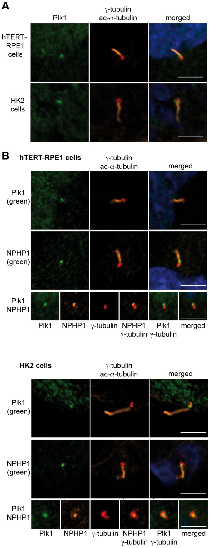
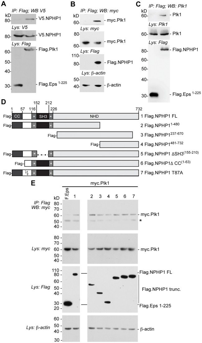
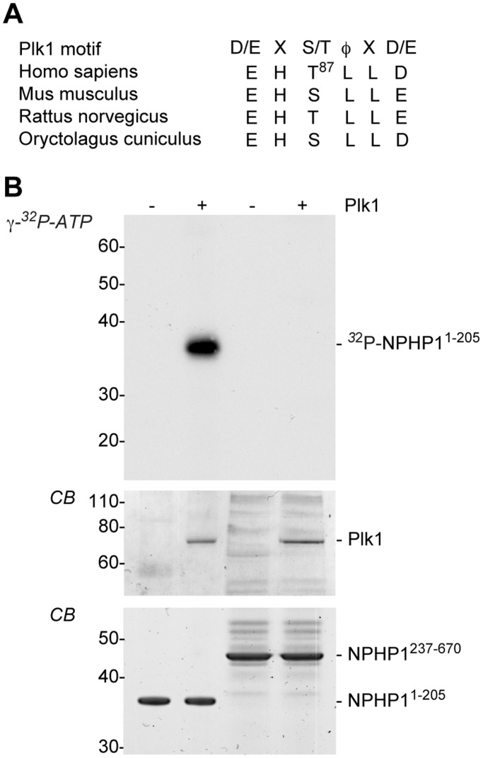
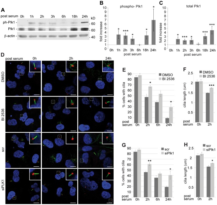
Polo-like kinase (Plk1) plays a central role in regulating the cell cycle. Plk1-mediated phosphorylation is essential for centrosome maturation, and for numerous mitotic events. Although Plk1 localizes to multiple subcellular sites, a major site of action is the centrosomes, which supports mitotic functions in control of bipolar spindle formation. In G0 or G1 untransformed cells, the centriolar core of the centrosome differentiates into the basal body of the primary cilium. Primary cilia are antenna-like sensory organelles dynamically regulated during the cell cycle. Whether Plk1 has a role in ciliary biology has never been studied. Nephrocystin-1 (NPHP1) is a ciliary protein; loss of NPHP1 in humans causes nephronophthisis (NPH), an autosomal-recessive cystic kidney disease. We here demonstrate that Plk1 colocalizes with nephrocystin-1 to the transition zone of primary cilia in epithelial cells. Plk1 co-immunoprecipitates with NPHP1, suggesting it is part of the nephrocystin protein complex. We identified a candidate Plk1 phosphorylation motif (D/E-X-S/T-φ-X-D/E) in nephrocystin-1, and demonstrated in vitro that Plk1 phosphorylates the nephrocystin N-terminus, which includes the specific PLK1 phosphorylation motif. Further, induced disassembly of primary cilia rapidly evoked Plk1 kinase activity, while small molecule inhibition of Plk1 activity or RNAi-mediated downregulation of Plk1 limited the first and second phase of ciliary disassembly. These data identify Plk1 as a novel transition zone signaling protein, suggest a function of Plk1 in cilia dynamics, and link Plk1 to the pathogenesis of NPH and potentially other cystic kidney diseases.
Conflict of interest statement
Figures
Similar articles
-
PCM1 recruits Plk1 to the pericentriolar matrix to promote primary cilia disassembly before mitotic entry.J Cell Sci. 2013 Mar 15;126(Pt 6):1355-65. doi: 10.1242/jcs.114918. Epub 2013 Jan 23. J Cell Sci. 2013. PMID: 23345402
-
DAZ-interacting Protein 1 (Dzip1) Phosphorylation by Polo-like Kinase 1 (Plk1) Regulates the Centriolar Satellite Localization of the BBSome Protein during the Cell Cycle.J Biol Chem. 2017 Jan 27;292(4):1351-1360. doi: 10.1074/jbc.M116.765438. Epub 2016 Dec 15. J Biol Chem. 2017. PMID: 27979967 Free PMC article.
-
Ubiquitylation-dependent localization of PLK1 in mitosis.Nat Cell Biol. 2013 Apr;15(4):430-9. doi: 10.1038/ncb2695. Epub 2013 Mar 3. Nat Cell Biol. 2013. PMID: 23455478 Free PMC article.
-
Mechanism of ciliary disassembly.Cell Mol Life Sci. 2016 May;73(9):1787-802. doi: 10.1007/s00018-016-2148-7. Epub 2016 Feb 11. Cell Mol Life Sci. 2016. PMID: 26869233 Free PMC article. Review.
-
Cell cycle progression by the repression of primary cilia formation in proliferating cells.Cell Mol Life Sci. 2013 Oct;70(20):3893-905. doi: 10.1007/s00018-013-1302-8. Epub 2013 Mar 9. Cell Mol Life Sci. 2013. PMID: 23475109 Free PMC article. Review.
Cited by
-
Oncoprotein CIP2A promotes the disassembly of primary cilia and inhibits glycolytic metabolism.EMBO Rep. 2018 May;19(5):e45144. doi: 10.15252/embr.201745144. Epub 2018 Feb 28. EMBO Rep. 2018. PMID: 29491003 Free PMC article.
-
The Role of Primary Cilia in Thyroid Cancer: From Basic Research to Clinical Applications.Front Endocrinol (Lausanne). 2021 Jun 8;12:685228. doi: 10.3389/fendo.2021.685228. eCollection 2021. Front Endocrinol (Lausanne). 2021. PMID: 34168619 Free PMC article. Review.
-
Activation of the oncogenic transcription factor B-Myb via multisite phosphorylation and prolyl cis/trans isomerization.Nucleic Acids Res. 2019 Jan 10;47(1):103-121. doi: 10.1093/nar/gky935. Nucleic Acids Res. 2019. PMID: 30321399 Free PMC article.
-
Cell Cycle Regulation of the Centrosome and Cilium.Drug Discov Today Dis Mech. 2013 Dec 1;10(3-4):e119-e124. doi: 10.1016/j.ddmec.2013.03.002. Drug Discov Today Dis Mech. 2013. PMID: 24982683 Free PMC article.
-
Cilia and coordination of signaling networks during heart development.Organogenesis. 2014 Jan 1;10(1):108-25. doi: 10.4161/org.27483. Epub 2013 Dec 17. Organogenesis. 2014. PMID: 24345806 Free PMC article. Review.
References
-
- Wheatley DN, Wang AM, Strugnell GE. Expression of primary cilia in mammalian cells. Cell Biol Int. 1996;20:73–81. - PubMed
Publication types
MeSH terms
Substances
Grants and funding
LinkOut - more resources
Full Text Sources
Molecular Biology Databases
Miscellaneous
