. 2012;7(6):e38839.
doi: 10.1371/journal.pone.0038839. Epub 2012 Jun 11.

Antioxidant protects against increases in low molecular weight hyaluronan and inflammation in asphyxiated newborn pigs resuscitated with 100% oxygen

Affiliations

Antioxidant protects against increases in low molecular weight hyaluronan and inflammation in asphyxiated newborn pigs resuscitated with 100% oxygen

Helene C D Østerholt et al. PLoS One. 2012.

Abstract

Background: Newborn resuscitation with 100% oxygen is associated with oxidative-nitrative stresses and inflammation. The mechanisms are unclear. Hyaluronan (HA) is fragmented to low molecular weight (LMW) by oxidative-nitrative stresses and can promote inflammation. We examined the effects of 100% oxygen resuscitation and treatment with the antioxidant, N-acetylcysteine (NAC), on lung 3-nitrotyrosine (3-NT), LMW HA, inflammation, TNFα and IL1ß in a newborn pig model of resuscitation.

Methods & principal findings: Newborn pigs (n = 40) were subjected to severe asphyxia, followed by 30 min ventilation with either 21% or 100% oxygen, and were observed for the subsequent 150 minutes in 21% oxygen. One 100% oxygen group was treated with NAC. Serum, bronchoalveolar lavage (BAL), lung sections, and lung tissue were obtained. Asphyxia resulted in profound hypoxia, hypercarbia and metabolic acidosis. In controls, HA staining was in airway subepithelial matrix and no 3-NT staining was seen. At the end of asphyxia, lavage HA decreased, whereas serum HA increased. At 150 minutes after resuscitation, exposure to 100% oxygen was associated with significantly higher BAL HA, increased 3NT staining, and increased fragmentation of lung HA. Lung neutrophil and macrophage contents, and serum TNFα and IL1ß were higher in animals with LMW than those with HMW HA in the lung. Treatment of 100% oxygen animals with NAC blocked nitrative stress, preserved HMW HA, and decreased inflammation. In vitro, peroxynitrite was able to fragment HA, and macrophages stimulated with LMW HA increased TNFα and IL1ß expression.

Conclusions & significance: Compared to 21%, resuscitation with 100% oxygen resulted in increased peroxynitrite, fragmentation of HA, inflammation, as well as TNFα and IL1ß expression. Antioxidant treatment prevented the expression of peroxynitrite, the degradation of HA, and also blocked increases in inflammation and inflammatory cytokines. These findings provide insight into potential mechanisms by which exposure to hyperoxia results in systemic inflammation.


Conflict of interest statement

Competing Interests: For this study a research grant award to MHW and RCS came from Ikaria Therapeutics. There are no patents, products in development or marketed products to declare. This does not alter the authors' adherence to all the PLoS ONE policies on sharing data and materials, as detailed online in the guide for authors.

Figures

Figure 1
Figure 1. Experimental protocol.
After anesthesia, ventilation, and instrumentation followed by 60 min of stabilization, pigs were subjected to asphyxia, followed by either 21% or 100% O2 resuscitation for 30 min, and then observed for a further 150 min after resuscitation when all animals were maintained in 21% O2. A separate group of animals resuscitated for 30 minutes with 100% O2 were treated with the antioxidant N-acetylcysteine (NAC) from the time of the start of resuscitation until the end of the experiment. Samples were obtained from five groups: Control animals were euthanized after undergoing surgical preparation and anesthesia but prior to asphyxia; the asphyxia group was harvested at the end of asphyxia prior to any resuscitation; and three groups of animals resuscitated with either 21% or 100% O2 or with 100% O2 and given NAC were harvested at 150 min after respective resuscitation strategies.
Figure 2
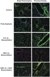
Figure 2. Dual immunostaining for 3-nitrotyrosine and HA.
Formalin-fixed, paraffin-embedded sections of inflated lungs were processed for dual label immunofluorescence using an antibody specific for 3-NT to localize peroxynitrite (red), and a biotinylated HA-binding probe to localize HA (green), with DAPI to label nuclei (blue). Control animals had abundant staining for HA in the sub-epithelial matrix around bronchiolar smooth muscle and on the endothelium of blood vessels, with less evident staining in the distal parenchyma and alveoli. At the end of asphyxia, there appeared to be a slight, but generalized decrease in HA staining in both the proximal airway as well as the distal parenchyma. Animals examined 150 minutes after 21% O2 resuscitation had HA staining that was not distinguishable from control animals, and had no staining for 3-NT. However, 150 minutes after 100% O2 exposure, little to no HA staining and an increase in 3-NT staining was observed throughout the lung. Animals resuscitated with 100% O2 and given NAC had little 3-NT staining and had HA staining similar to that of animals resuscitated with 21% O2.
Figure 3
Figure 3. Hyaluronan content in bronchoalveolar lavage and serum, and expression of enzymes regulating hyaluronan synthesis and degradation.
HA content was determined in BAL (A) and in serum (B). At the end of asphyxia, the HA content of BAL decreased and that in the serum increased. Resuscitation with 21% O2 did not increase HA either in the BAL or in the serum. However, BAL HA was significantly increased in animals resuscitated with 100% O2, and this increase was completely inhibited by treatment with NAC (A). The changes in hyaluronan synthase 1 (has1), has2 and has3, as well as hyaluronidase 1 (hyal1) and 2 (hyal2) were determined using quantitative RT-PCR. There were no changes in has1 and has 3 expression (data not shown). Expression of has2 (C) showed a trend to increased expression after asphyxia and 21% O2 resuscitation. However, treatment with NAC significantly inhibited has2 expression compared to 100% O2 resuscitation alone (C). The expression of hyal1 (D) and hyal2 (E) remained unchanged except that 100% O2 exposed animals treated with NAC showed significantly increased expression of both hyaluronidases.
Figure 4
Figure 4. HA size determination.
HA size was determined in lung tissue and two representative samples per group are shown in this gel. Control animals had HMW HA and asphyxia was associated with some degradation of HA. Resuscitation with 21% O2 was associated with HMW HA where as resuscitation with 100% O2 showed marked degradation of HA. Interestingly, treatment of 100% O2 resuscitated animals with NAC was associated with a preservation of HMW HA.
Figure 5
Figure 5. Lung myeloperoxidase and N-acetylglucosaminidase activities as measures of neutrophil and macrophage contents respectively.
Macrophage content was determined by NAG activity and neutrophil content was determined by MPO activity as described previously and in Materials and Methods. Data were segregated according to the molecular size of HA found in the lung and plotted as box and whisker plots with outliers shown as additional dots. Both NAG (A) and MPO (B) activities were significantly higher in animals that had LMW HA. Newborn pigs resuscitated with 100% O2 and treated with NAC had significantly lower NAG (C) and MPO (D) activities than those without NAC treatment, suggesting that antioxidant treatment decreases the inflammatory response to resuscitation with hyperoxia.
Figure 6
Figure 6. Changes in BAL TNFα and IL1ß concentrations.
The BAL contents of TNFα (A) and IL1ß (B) were determined by ELISA were also segregated according to the molecular size of HA. Data are presented as box and whisker plots of 25th and 75th percentiles showing outliers as additional dots. Both inflammatory markers were significantly higher in the animals that had LMW HA in the lung. Newborn pigs resuscitated with 100% O2 and treated with NAC had significantly lower TNFα (C) and IL1ß (D) than those without NAC treatment, suggesting that antioxidant treatment decreases the inflammatory response to resuscitation with hyperoxia.
Figure 7
Figure 7. Fragmentation of HA by peroxynitrite and stimulation of TNFα and IL1ß by oligomeric HA in vitro.
HMW HA (Healon™, 1×10⁶ Da, Lane 2) was exposed to 100 µM 3-morpholinosydnonimine (SIN-1), a compound that spontaneously releases NO and superoxide to generate peroxynitrite. Exposure of HMW HA to SIN-1 results in degradation to LMW HA (300–500 kDa, Lane 6). Exposure to SIN-1 in the presence of 600 mU/ml superoxide dismutase (SOD) partially protects this degradation (Lane 7). Exposure to 300 µM PAPANOATE, a pure NO donor, does not degrade Healon™ (Lane 8). In addition, treatment of Healon™ with either 1 U/ml Streptomyces hyaluronidase at 60°C for 2 hours (Lane 4) or sonication for 2 minutes (Lane 5) results in formation of LMW HA. Just heating HMW HA at 60°C in the absence of hyaluronidase did not degrade HA in the same manner as in the presence of enzyme (Lane 2). Lane 1 has 200 kDa HA (ICN) and Lane 9 contains Hind III digested DNA makers.
Figure 8
Figure 8. Stimulation of TNFα and IL1ß production by a 6mer HA oligosaccharide.
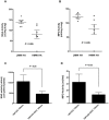
RAW264.7 murine macrophages (1×10⁶) were stimulated with a 10 µg/ml of 6-mer HA oligosaccharide in vitro. Supernatant IL1ß and TNFα were measured using standard ELISA. HA6 significantly stimulated the expression of both IL1ß and TNFα in the macrophage cell line, thereby confirming that LMW HA can stimulate the expression of inflammatory cytokines in macrophages.
Figure 9
Figure 9. Overall model of the role of oxidative and nitrative stresses and LMW HA in asphyxia and hyperoxia-stimulated inflammation.
Exposure to 100% oxygen in asphyxiated newborn pigs results in the production of superoxide and peroxynitrite that cause the fragmentation of HMW to LMW HA. LMW HA, in turn stimulates inflammatory cytokine expression in macrophages and promotes inflammation. Strategies to prevent the formation of LMW HA, such as limiting oxygen exposure or treatment with antioxidants (the current work) will result in decreased inflammation. This model predicts that direct blockade of LMW HA should also achieve the same result.

References

    1. Bryce J, Boschi-Pinto C, Shibuya K, Black RE. WHO estimates of the causes of death in children. Lancet. 2005;365:1147–1152. - PubMed
    1. Alonso-Spilsbury M, Mota-Rojas D, Villanueva-Garcia D, Martinez-Burnes J, Orozco H. Perinatal asphyxia pathophysiology in pig and human: a review. Animal reproduction science. 2005;90:1–30. - PubMed
    1. Obladen M. History of neonatal resuscitation. Part 2: oxygen and other drugs. Neonatology. 2009;95:91–96. - PubMed
    1. Saugstad OD, Ramji S, Soll RF, Vento M. Resuscitation of newborn infants with 21% or 100% oxygen: an updated systematic review and meta-analysis. Neonatology. 2008;94:176–182. - PubMed
    1. Richmond S, Goldsmith JP. Refining the role of oxygen administration during delivery room resuscitation: what are the future goals? Seminars in fetal & neonatal medicine. 2008;13:368–374. - PubMed

Publication types

MeSH terms