Review
. 2012 Oct;272(4):317-29.
doi: 10.1111/j.1365-2796.2012.02564.x. Epub 2012 Jul 29.

New insights into osteoporosis: the bone-fat connection

Affiliations
Review

New insights into osteoporosis: the bone-fat connection

M Kawai et al. J Intern Med. 2012 Oct.

Abstract

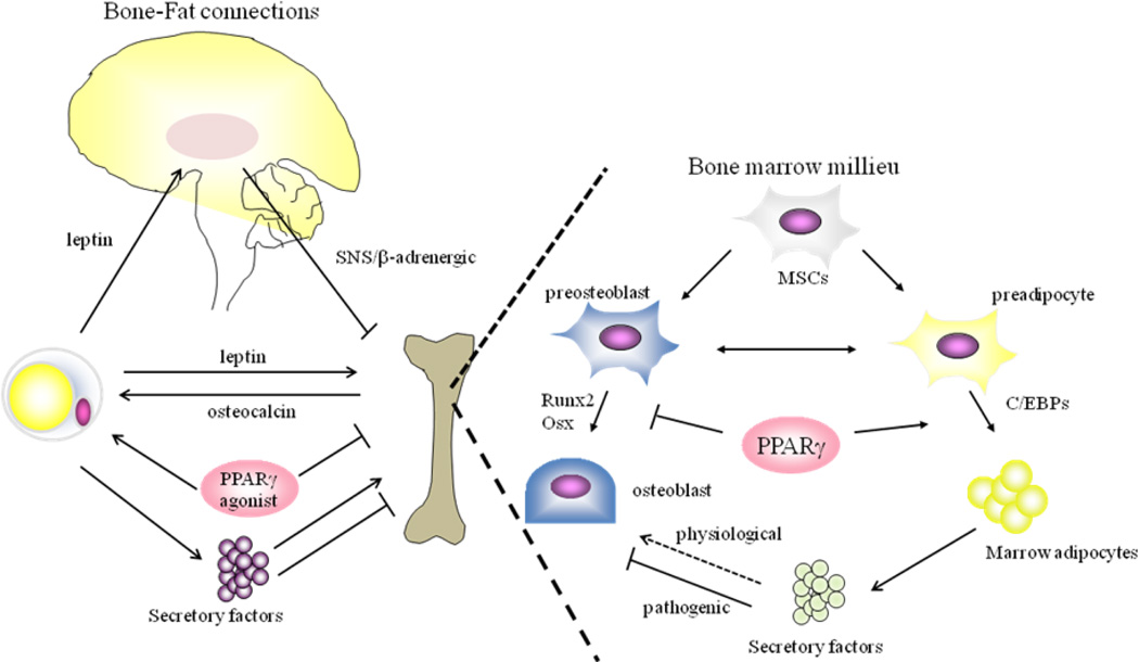
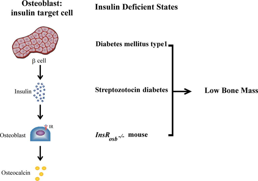
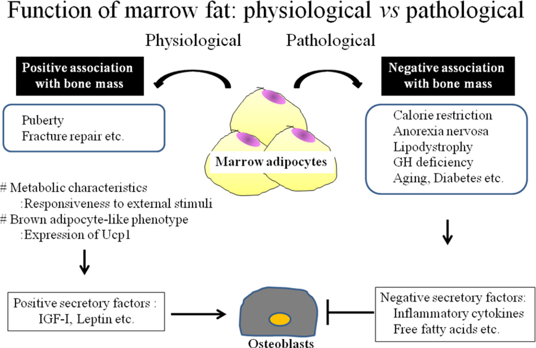
Osteoporosis and obesity are chronic disorders that are both increasing in prevalence. The pathophysiology of these conditions is multifactorial and includes genetic, environmental and hormonal determinants. Although it has long been considered that these are distinct disorders rarely found in the same individual, emerging evidence from basic and clinical studies support an important interaction between adipose tissue and the skeleton. It is proposed that adiposity may influence bone remodelling through three mechanisms: (i) secretion of cytokines that directly target bone, (ii) production of adipokines that influence the central nervous system thereby changing sympathetic impulses to bone and (iii) paracrine influences on adjacent skeletal cells. Here we focus on the current understanding of bone-fat interactions and the clinical implications of recent studies linking obesity to osteoporosis.


Figures

Figure 1
Figure 1. Schematic model of Bone-Fat connections
Leptin regulation of bone mass includes: 1- activation of SNS/β-adrenergic signaling through hypothalamic integration, which results in bone loss, 2- direct anabolic effects on osteoblasts, although the precise mechanisms of leptin action on skeletal homeostasis remain to be elucidated. Osteocalcin produced by osteoblasts decreases fat mass in part through promoting adiponectin production in adipocytes and activation of PPARγ has been shown to cause increased adipose tissue mass and bone loss. Secretory factors produced from adipocytes, such as imflammatory cytokines, fatty acid, leptin and adiponectin, can positively or negatively regulate skeletal mass. In a bone marrow milieu, marrow adipocytes play a pivotal role in the regulation of osteoblast function. Secretory factors including inflammatory cytokines are also produced by marrow adipocytes. These cytokines are possibly acting on osteoblasts in a paracrine manner and suppressing osteoblast function and/or differentiation in pathogenic conditions, whereas in physiological conditions, marrow adipocytes could be positively regulating bone mass, but this concept needs to be clarified. In addition, activation of PPARγ causes bone loss in part through altering the specification of MSCs towards adipogenesis and away from osteogenesis, resulting in the decreased osteoblast pool in the bone marrow microenvironment. SNS: sympathetic nervous system, PPAR: peroxisome proliferative activated receptors, C/EBP: CCAAT/enhancer binding proteins; RUNX: runt-related transcription factor, MSC: mesenchymal stem cell,
Figure 2
Figure 2
Insulin regulates skeletal homeostasis in part by stimulating new bone formation and bone remodeling. Insulin deficiency or resistance leads to low bone mass and increased skeletal fragility. Enhanced bone resorption increases release of undercarboxylated osteocalcin which in turn can enhance insulin secretion and adipocyte sensitivity(7)
Figure 3
Figure 3. Function of marrow fat
Marrow adipocytes produce a number of secretory factors. Such factors could have a significant role in osteoblast differentiation and/or function. In pathogenic conditions, these determinants could impact osteoblasts in a negative direction, whereas in physiological conditions these factors may have a different role from the one observed in the pathogenic conditions. There is also evidence that marrow fat is metabolically active and that genes which are characteristics to brown adipocytes are expressed in marrow adipocytes.

Similar articles

Cited by

References

    1. Karsenty G. Convergence between bone and energy homeostases: leptin regulation of bone mass. Cell Metab. 2006;4:341–348. - PubMed
    1. Friedman JM. Leptin at 14 y of age: an ongoing story. Am J Clin Nutr. 2009;89:973S–979S. - PMC - PubMed
    1. Ryden M, Arner P. Tumour necrosis factor-alpha in human adipose tissue -- from signalling mechanisms to clinical implications. J Intern Med. 2007;262:431–438. - PubMed
    1. Langsted A, Freiberg JJ, Tybjaerg-Hansen A, Schnohr P, Jensen GB, Nordestgaard BG. Nonfasting cholesterol and triglycerides and association with risk of myocardial infarction and total mortality: the Copenhagen City Heart Study with 31 years of follow-up. J Intern Med. 2011;270:65–75. - PubMed
    1. Garg A. Clinical review#: Lipodystrophies: genetic and acquired body fat disorders. J Clin Endocrinol Metab. 2011;96:3313–3325. - PMC - PubMed