. 2012 May-Jun;19(3):196-206.
doi: 10.1111/j.1399-3089.2012.00705.x.

Complete absence of the αGal xenoantigen and isoglobotrihexosylceramide in α1,3galactosyltransferase knock-out pigs

Affiliations

Complete absence of the αGal xenoantigen and isoglobotrihexosylceramide in α1,3galactosyltransferase knock-out pigs

Gisella L Puga Yung et al. Xenotransplantation. 2012 May-Jun.

Abstract

Background: Anti-Galα1,3Galβ-R natural antibodies are responsible for hyperacute rejection in pig-to-primate xenotransplantation. Although the generation of pigs lacking the α1,3galactosyltransferase (GalT) has overcome hyperacute rejection, antibody-mediated rejection is still a problem. It is possible that other enzymes synthesize antigens similar to Galα1,3Gal epitopes that are recognized by xenoreactive antibodies. The glycosphingolipid isoglobotrihexosylceramide (iGb₃) represents such a candidate expressing an alternative Galα1,3Gal epitope. The present work determined whether the terminal Galα1,3Gal disaccharide is completely absent in Immerge pigs lacking the GalT using several different highly sensitive methods.

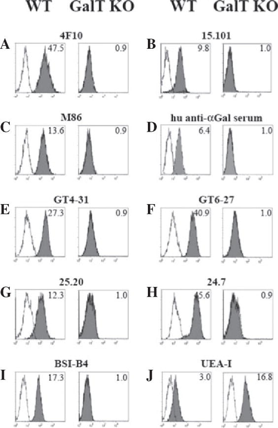
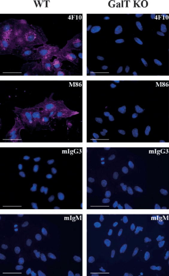
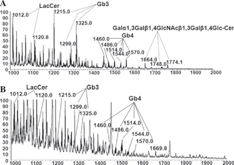
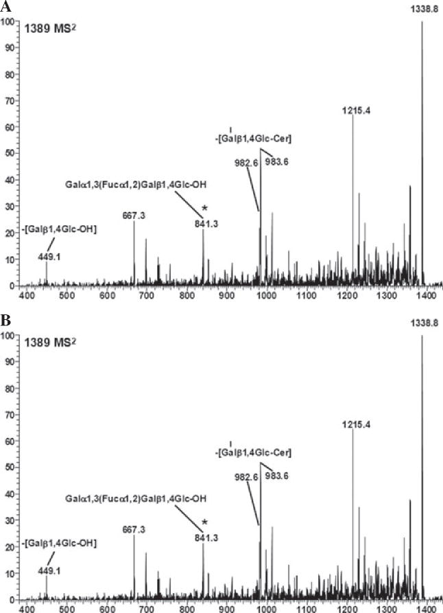
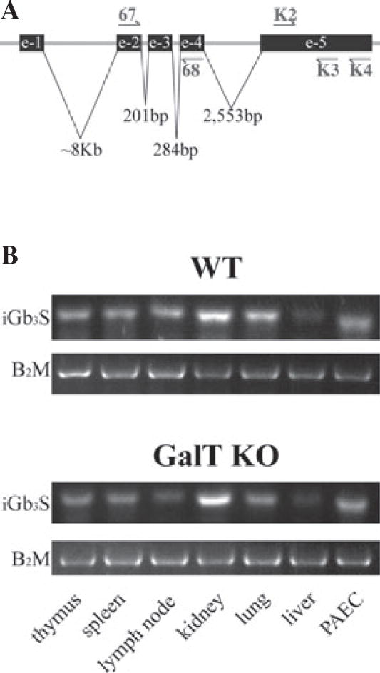
Methods: The expression of Galα1,3Gal was evaluated using a panel of antibodies and lectins by flow cytometry and fluorescent microscopy; GalT activity was detected by an enzymatic assay; and ion trap mass spectroscopy of neutral cellular membranes extracted from aortic endothelial was used for the detection of sugar structures. Finally, the presence of iGb₃ synthase mRNA was tested by RT-PCR in pig thymus, spleen, lymph node, kidney, lung, and liver tissue samples.

Results: Aortic endothelial cells derived from GalT knockout pigs expressed neither Galα1,3Gal nor iGb₃ on their surface, and GalT enzymatic activity was also absent. Lectin staining showed an increase in the blood group H-type sugar structures present in GalT knockout cells as compared to wild-type pig aortic endothelial cells (PAEC). Mass spectroscopic analysis did not reveal Galα1,3Gal in membranes of GalT knockout PAEC; iGb₃ was also totally absent, whereas a fucosylated form of iGb₃ was detected at low levels in both pig aortic endothelial cell extracts. Isoglobotrihexosylceramide 3 synthase mRNA was expressed in all pig tissues tested whether derived from wild-type or GalT knockout animals.

Conclusions: These results confirm unequivocally the absence of terminal Galα1,3Gal disaccharides in GalT knockout endothelial cells. Future work will have to focus on other mechanisms responsible for xenograft rejection, in particular non-Galα1,3Gal antibodies and cellular responses.


Conflict of interest statement

Conflict of interest

DZ is a consultant for BioTex, Houston, TX, and an inventor involved in patents related to technologies mentioned in this article, issued or in application.

Figures

Fig. 1
Fig. 1
Lack of iGb3 and αGal expression on pig aortic endothelial cells derived from α1,3galactosyltransferase knockout pigs analyzed by flow cytometry. The epitopes iGb3 and αGal were tested in PAEC cells by indirect flow cytometry. Two mAb recognizing iGb3: clone 4F10 (A) with specificity for both αGal and iGb3 and clone 15.101 (B) were assayed in addition to the commercial mAb clone M86 (C); polyclonal purified anti-αGal from human serum (D); others mAb anti-αGal as the clone GT4-31 (E); clone GT6-27 (F); clone 25.20 (G); and clone 24.7 (H). Lectin staining with BSI-B4 (I) which binds to terminal α-D-galactosyl residues present in αGal and the UEA-I (J) which has with affinity for L-Fuc are also shown. Gray-shaded histograms show the staining for the different mAb, human serum, and lectins. Open histograms show the matching isotype controls when mAb were tested, human anti-αGal depleted serum for human serum, and cells incubated only with staining buffer for lectins staining. MFIR are shown in the upper right corner. Representative experiments from 3 to 5 different stainings are shown. BSI-B4, Bandeiraea simplicifolia lectin; GalT KO, α1,3galactosyltransferase knockout; iGb3, isoglobotrihexosylceramide 3; mAb, monoclonal antibodies; MFIR, Mean fluorescence intensity ratio; PAEC, pig aortic endothelial cell; UEA-I, Ulex europaeus lectin.
Fig. 2
Fig. 2
Uniform distribution of αGal in wild-type pig aortic endothelial cells. PAEC WT (left column) and GalT KO (right column) were grown directly in 96-well plates; fixed/permeabilized before staining with the 4F10 and M86 mAb or matching isotype controls: IgM and IgG3, respectively, as indicated in the top-right corner of the figures; and analyzed by Olympus fluorescent IX71 microscope fluorescent microscope. Overlay pictures of Evans blue channel in violet and DAPI channel in blue. Bar corresponds to 50 μm. Representative images of four different staining with the different pig cell lines. GalT KO, α1,3galactosyltransferase knockout; mAb, monoclonal antibodies; PAEC, pig aortic endothelial cell; WT, wild-type.
Fig. 3
Fig. 3
Absence of αGal and iGb3 in pig aortic endothelial cell derived from α1,3galactosyltransferase knockout pigs analyzed by electrospray ionization mass spectroscopy. Neutral glycosphingolipids membranes from PAEC were extracted, permethylated, and analyzed by MS. MS was carried out in positive ion mode on a linear ion trap mass spectrometer using a nano-electrospray source. MS1 neutral glycolipid fractions of WT (A) and GalT KO (B) PAEC were extracted and analyzed. Molecular ions containing αGal epitope (Galα3nLc4, Galα3Galβ4GlcNAcβ3Galβ4Glc-Cer) are only seen in WT PAEC (1664; 1748; and 1774 m/z). The molecular ions representing regioisomers of trihexosyceramides (Gb3/iGb3, 1215, 1299, and 1325 m/z) present in PAEC were further subjected to MS4 analysis by ion trap mass spectrometry excluding the presence of iGb3 [17]. The Gb3/iGb3 precursor, LacCer (1012 and 1120 m/z), is present in both PAEC Spectra correspond to x-axe in m/z and y-axe relative absorbance. Gb3, globotriaosylceramide also known as Pk antigen; iGb3, isoglobotrihexosylceramide; LacCer, lactosylceramide; PAEC, pig aortic endothelial cells; MS, mass spectroscopy.
Fig. 4
Fig. 4
Wild-type and α1,3galactosyltransferase knockout neutral pig aortic endothelial cell membranes contain a fucosylated form of iGb3. Neutral glycosphingolipids membranes from WT and GalT KO PAEC were extracted, permethylated, and analyzed by MS. Molecular ion profiles of neutral glycolipid membrane fractions of WT (A) and GalT KO (B) PAEC obtained by linear ion trap mass spectrometer using the electrospray ionization mass spectroscopic method (LTQ-ESI-MS) are shown. The presence of fucosylated iGb3 structure was found by precursor ion mapping method (using 841 as the product ion). Candidate structures for the ion are Galα1,3(Fucα1,2) Galβ1,4Glc-Cer, and/or Fucα1,2Galα1,3Galβ1,4Glc-Cer. Spectra x-axe in m/z. Cer, ceramide; GalT KO, α1,3galactosyltransferase knockout; Gb3, globotrihexosylceramide 3, iGb3, isoglobotrihexo sylceramide; MS, mass spectrometry; PAEC, pig aortic endothelial cells; WT, wild-type.
Fig. 5
Fig. 5
Expression of mRNA levels of the isoglobotrihexosylceramide 3 synthase in pig aortic endothelial cells and pig tissues. Different tissues were isolated from just sacrificed animals and snap frozen for RNA extraction and analysis. The mRNA levels for iGb3S were checked with three different set of primers. Location of primers is shown using the patented WO 2005/047469 sequence as reference (A). Pig β2-microglobulin was used as a housekeeping gene. RT-PCR products of K2/K3 primers set were run in 2% agarose gel electrophoresis (B). Two different samples from each organ were obtained from a WT animal (#16517, female, AA haplotype, 2.5 yr old) and a GalT KO animal (#16183, female, DD haplotype, 3.5 yr old), respectively. e, exon; GalT KO, α1,3galactosyltransferase knockout; iGb3S, isoglobotrihexosylceramide 3 synthase; mRNA, messenger ribonucleic acid; PAEC, pig aortic - endothelial cells.

Similar articles

Cited by

References

    1. Tai HC, Zhu X, Hara H, et al. The pig-to-primate immune response: relevance for xenotransplantation. Xenotransplantation. 2007;14:227–235. - PubMed
    1. Li S, Waer M, Billiau AD. Xenotransplantation: role of natural immunity. Transpl Immunol. 2009;21:70–74. - PubMed
    1. Macher BA, Galili U. The Galalpha1,3Galbeta1,4Glc-NAc-R (alpha-Gal) epitope: a carbohydrate of unique evolution and clinical relevance. Biochim Biophys Acta. 2008;1780:75–88. - PMC - PubMed
    1. Galili U, Shohet SB, Kobrin E, Stults CL, Macher BA. Man, apes, and Old World monkeys differ from other mammals in the expression of alpha-galactosyl epitopes on nucleated cells. J Biol Chem. 1988;263:17755–17762. - PubMed
    1. Hisashi Y, Yamada K, Kuwaki K, et al. Rejection of cardiac xenografts transplanted from a 1,3-galactosyltransferase gene-knockout (GalT-KO) pigs to baboons. Am J Transplant. 2008;8:2516–2526. - PMC - PubMed

Publication types

MeSH terms