. 2013 Apr;33(2):122-33.
doi: 10.1111/j.1440-1789.2012.01332.x. Epub 2012 Jun 18.

Frontotemporal lobar degeneration with TDP-43 proteinopathy and chromosome 9p repeat expansion in C9ORF72: clinicopathologic correlation

Affiliations

Frontotemporal lobar degeneration with TDP-43 proteinopathy and chromosome 9p repeat expansion in C9ORF72: clinicopathologic correlation

Eileen H Bigio et al. Neuropathology. 2013 Apr.

Abstract

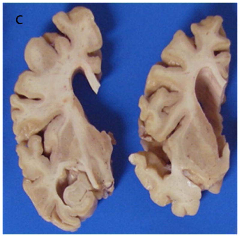
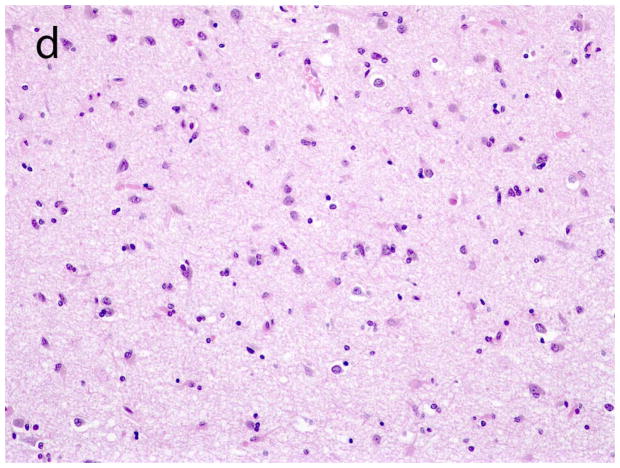
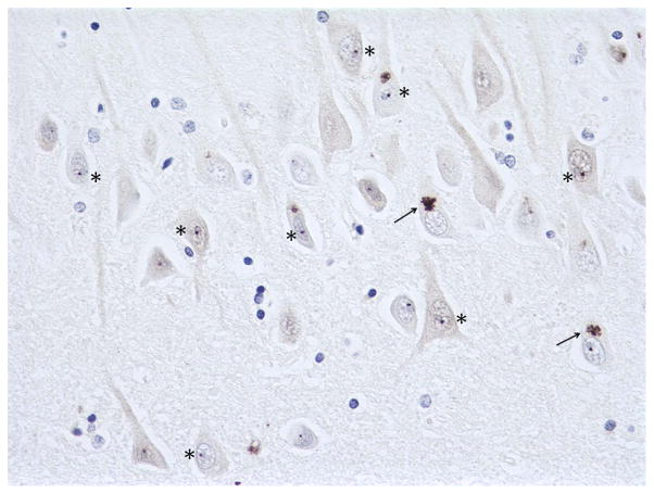
Mutations in C9ORF72 resulting in expanded hexanucleotide repeats were recently reported to be the underlying genetic abnormality in chromosome 9p-linked frontotemporal lobar degeneration with TAR DNA-binding protein of 43 kD (TDP-43) proteinopathy (FTLD-TDP), amyotrophic lateral sclerosis (ALS), and frontotemporal lobar degeneration with motor neuron disease (FTLD-MND). Several subsequent publications described the neuropathology as being similar to that of FTLD-TDP and ALS without C9ORF72 mutations, except that cases with mutations have p62 and ubiquitin positive, TDP-43 negative inclusions in cerebellum, hippocampus, neocortex, and basal ganglia. The identity of this protein is as yet unknown, and its significance is unclear. With the goal of potentially uncovering the significance of these inclusions, we compared the clinical, pathologic and genetic characteristics in cases with C9ORF72 mutations to those without. We confirmed the apparent specificity of p62 positive, TDP-43 negative inclusions to cases with C9ORF72 mutations. In hippocampus, these inclusions correlated with hippocampal atrophy. No additional correlations were uncovered. However, this is the first report to show that although most cases with C9ORF72 mutations were TDP type B, some of the pathologic characteristics in these cases were more similar to TDP types A and C than to type B cases. These include greater cortical and hippocampal atrophy, greater ventricular dilatation, more neuronal loss and gliosis in temporal lobe and striatum, and TDP-43 positive fine neuritic profiles in the hippocampus, implying that the C9ORF72 mutation modifies the pathologic phenotype of FTLD-TDP type B.


Figures

Figure 1
Figure 1
Examples of varying degrees of gross cortical atrophy. Severe atrophy of the posterior portions of the superior frontal gyrus (sfg) and middle frontal gyrus (mfg) and of the entire motor cortex (m), moderate atrophy of the frontal pole and the posterior portion of the superior temporal gyrus (stg), mild atrophy of the remainder of the frontal and temporal cortex, and no appreciable atrophy of the parietal cortex. Note: Moderate gross atrophy of the frontal pole of case 4 is shown in Figure 3 of reference #.
Figure 2
Figure 2
Examples of varying degrees of ventricular dilatation. Mild (a), moderate (b), and severe (c) ventricular dilatation.
Figure 2
Figure 2
Examples of varying degrees of ventricular dilatation. Mild (a), moderate (b), and severe (c) ventricular dilatation.
Figure 2
Figure 2
Examples of varying degrees of ventricular dilatation. Mild (a), moderate (b), and severe (c) ventricular dilatation.
Figure 3
Figure 3
Examples of cortical neuronal loss and gliosis. Cortex with no (a), mild (b), moderate (c), and severe (d) neuronal loss and gliosis.
Figure 3
Figure 3
Examples of cortical neuronal loss and gliosis. Cortex with no (a), mild (b), moderate (c), and severe (d) neuronal loss and gliosis.
Figure 3
Figure 3
Examples of cortical neuronal loss and gliosis. Cortex with no (a), mild (b), moderate (c), and severe (d) neuronal loss and gliosis.
Figure 3
Figure 3
Examples of cortical neuronal loss and gliosis. Cortex with no (a), mild (b), moderate (c), and severe (d) neuronal loss and gliosis.
Figure 4
Figure 4
Dentate gyrus cytoplasmic inclusions. Sparse (case 3) (a) and moderate (case 1) (b) TDP-43 positive neuronal cytoplasmic inclusions in the dentate gyrus. (both 400x).
Figure 5
Figure 5
Hippocampal dentate gyrus of case 5 with frequent cytoplasmic inclusions labeled with p62 (a) and with TDP-43 (b) (both 400x). Note that fewer are labeled with TDP-43 than with p62.
Figure 6
Figure 6
Synaptic TDP-43 labeling, CA2-4 region of the hippocampus. Sparse (case 5, CA2-3 region) (a) and moderate (case 4, CA3-4 region) (b) synaptic labeling in CA2-4 region of the hippocampus.
Figure 7
Figure 7
Frequent TDP-43 positive fine dystrophic neurites in CA1 of case 5 with C9ORF72 mutation (a) and case 10 without mutation, FTLD-TDP type A (b) (400x).
Figure 8
Figure 8
Cerebellar granular neurons with p62 positive cytoplasmic inclusions (case 2) (a); same field showing fewer ubiquitin positive cytoplasmic inclusions (b) (both 600x). A rare p62 positive intranuclear inclusion in a cerebellar granular neuron (arrow) (case 3) (c) (600x). Cerebellar molecular layer with p62 positive cytoplasmic inclusions in basket cells (arrows) and one Purkinje cell (asterisk) (case 3) (d) (200x).
Figure 9
Figure 9
Hippocampal CA3 region of case 2 showing several p62 positive cytoplasmic inclusions, some star-shaped (arrows), and numerous intranuclear dot-like inclusions (asterisks) (400x).
Figure 10
Figure 10
p62 positive star-shaped cytoplasmic inclusion in pyramidal neuron of frontal cortex of case 5 (600x).

References

    1. DeJesus-Hernandez M, Mackenzie IR, Boeve BF, et al. Expanded GGGGCC hexanucleotide repeat in non-coding region of C9ORF72 causes chromosome 9p-linked FTD and ALS. Neuron. 2011;72:245–256. - PMC - PubMed
    1. Renton AE, Majounie E, Waite A, et al. A nexanucleotide repeat expansion in C9ORF72 is the cause of chromosome 9p21-linked ALS-FTD. Neuron. 2011;72:257–268. - PMC - PubMed
    1. Gijselinck I, Van Langenhove T, van der Zee J, et al. A Cporf72 promoter repeat expansion in a Flanders-Belgian cohort with disorders of the frontotemporal lobar degeneration-amyotrophic lateral sclerosis spectrum: a gene identification study. Lancet Neurology. 2012;11:54–65. - PubMed
    1. Murray ME, DeJesus-Hernandez M, Rutherford N, et al. Clinical and neuropathologic heterogeneity of c9FTD/ALS associated with hexanucleotide repeat expansion in C9ORF72. Acta Neuropathol. 2011;122:673–690. - PMC - PubMed
    1. Al-Sarraj S, King A, Troakes C, et al. p62 positive, TDP-43 negative, neuronal cytoplasmic and intranuclear inclusions in the cerebellum and hippocampus define the pathology of C9orf72 linked FTLD and MND/ALS. Acta Neuropathol. 2011;122:691–702. - PubMed

Publication types