Review
. 2012:13:263-83.
doi: 10.1146/annurev-genom-090711-163729. Epub 2012 Jun 6.

The evolution of human genetic studies of cleft lip and cleft palate

Affiliations
Review

The evolution of human genetic studies of cleft lip and cleft palate

Mary L Marazita. Annu Rev Genomics Hum Genet. 2012.

Abstract

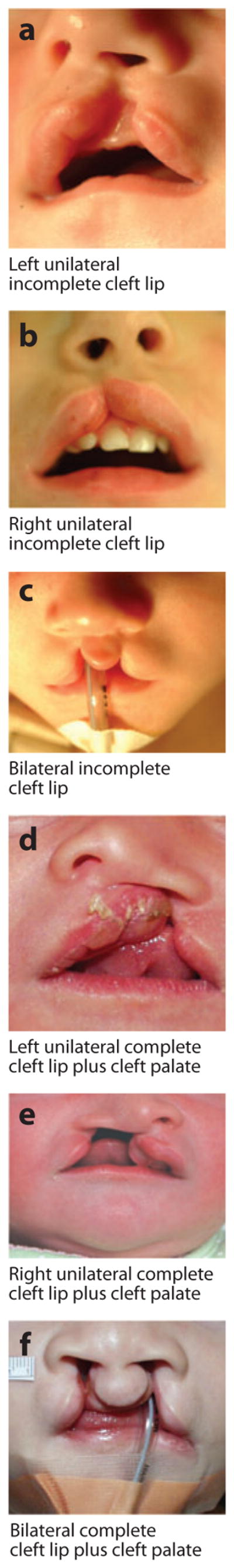
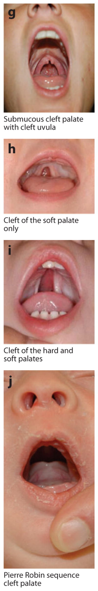
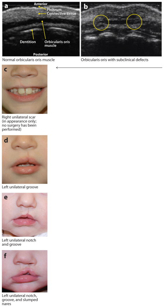
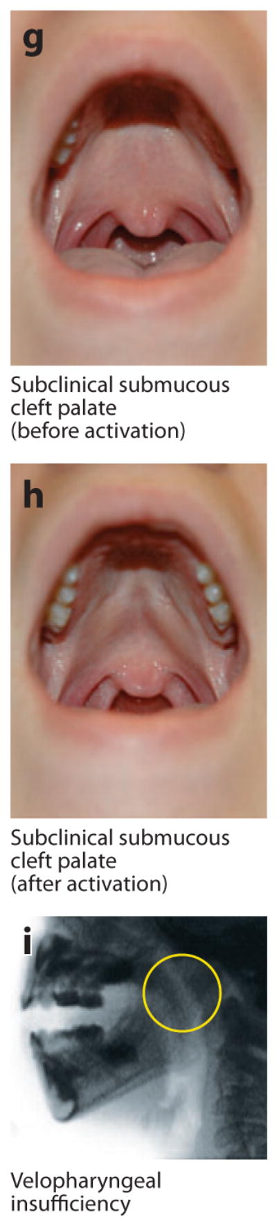
Orofacial clefts (OFCs)--primarily cleft lip and cleft palate--are among the most common birth defects in all populations worldwide, and have notable population, ethnicity, and gender differences in birth prevalence. Interest in these birth defects goes back centuries, as does formal scientific interest; scientists often used OFCs as examples or evidence during paradigm shifts in human genetics, and have also used virtually every new method of human genetic analysis to deepen our understanding of OFC. This review traces the evolution of human genetic investigations of OFC, highlights the specific insights gained about OFC through the years, and culminates in a review of recent key OFC genetic findings resulting from the powerful tools of the genomics era. Notably, OFC represents a major success for genome-wide approaches, and the field is poised for further breakthroughs in the near future.


Figures

Figure 1
Figure 1
Examples of overt types of orofacial clefts. Photographs courtesy of M. Ford, Children’s Hospital of Pittsburgh Cleft-Craniofacial Center, through FaceBase (http://www.facebase.org).
Figure 1
Figure 1
Examples of overt types of orofacial clefts. Photographs courtesy of M. Ford, Children’s Hospital of Pittsburgh Cleft-Craniofacial Center, through FaceBase (http://www.facebase.org).
Figure 2
Figure 2
Examples of microform and subclinical phenotypes. (a) Normal orbicularis oris muscle visualized via high-resolution ultrasound in a cross section through the upper lip. Note the wide, uniform appearance of the muscle and contrast with the breaks seen in panel b. (b) Orbicularis oris muscle with subclinical (i.e., not externally visible) defects that appear as breaks in the muscle. This image shows bilateral breaks (circled), which, notably, are located where overt clefts of the lip would be. (cf) Microform lip defects. (gh) Subclinical submucous cleft palate (i.e., not obvious unless activated by appropriate speech sounds; these panels show the same individual before and after activation). (i) Velopharyngeal insufficiency visualized with videofluoroscopy. Note the wide velopharyngeal gap (circled). Photographs in panels ci courtesy of M. Ford, Children’s Hospital of Pittsburgh Cleft-Craniofacial Center, through FaceBase (http://www.facebase.org).
Figure 2
Figure 2
Examples of microform and subclinical phenotypes. (a) Normal orbicularis oris muscle visualized via high-resolution ultrasound in a cross section through the upper lip. Note the wide, uniform appearance of the muscle and contrast with the breaks seen in panel b. (b) Orbicularis oris muscle with subclinical (i.e., not externally visible) defects that appear as breaks in the muscle. This image shows bilateral breaks (circled), which, notably, are located where overt clefts of the lip would be. (cf) Microform lip defects. (gh) Subclinical submucous cleft palate (i.e., not obvious unless activated by appropriate speech sounds; these panels show the same individual before and after activation). (i) Velopharyngeal insufficiency visualized with videofluoroscopy. Note the wide velopharyngeal gap (circled). Photographs in panels ci courtesy of M. Ford, Children’s Hospital of Pittsburgh Cleft-Craniofacial Center, through FaceBase (http://www.facebase.org).

References

    1. Ardinger HH, Buetow KH, Bell GI, Bardach J, VanDemark DR, Murray JC. Association of genetic variation of the transforming growth factor-alpha gene with cleft lip and palate. Am J Hum Genet. 1989;45:348–53. - PMC - PubMed
    1. Ashique AM, Fu K, Richman JM. Endogenous bone morphogenetic proteins regulate outgrowth and epithelial survival during avian lip fusion. Development. 2002;129:4647–60. - PubMed
    1. Baek JA, Lan Y, Liu H, Maltby KM, Mishina Y, Jiang R. Bmpr1a signaling plays critical roles in palatal shelf growth and palatal bone formation. Dev Biol. 2011;350:520–31. - PMC - PubMed
    1. Bakrania P, Efthymiou M, Klein JC, Salt A, Bunyan DJ, et al. Mutations in BMP4 cause eye, brain, and digit developmental anomalies: overlap between the BMP4 and hedgehog signaling pathways. Am J Hum Genet. 2008;82:304–19. - PMC - PubMed
    1. Baltimore D. Our genome unveiled. Nature. 2001;409:814–16. - PubMed

Publication types