Review
. 2012 Jul 24;51(29):5705-24.
doi: 10.1021/bi300752r. Epub 2012 Jul 9.

Mechanisms of mammalian iron homeostasis

Affiliations
Review

Mechanisms of mammalian iron homeostasis

Kostas Pantopoulos et al. Biochemistry. .

Abstract

Iron is vital for almost all organisms because of its ability to donate and accept electrons with relative ease. It serves as a cofactor for many proteins and enzymes necessary for oxygen and energy metabolism, as well as for several other essential processes. Mammalian cells utilize multiple mechanisms to acquire iron. Disruption of iron homeostasis is associated with various human diseases: iron deficiency resulting from defects in the acquisition or distribution of the metal causes anemia, whereas iron surfeit resulting from excessive iron absorption or defective utilization causes abnormal tissue iron deposition, leading to oxidative damage. Mammals utilize distinct mechanisms to regulate iron homeostasis at the systemic and cellular levels. These involve the hormone hepcidin and iron regulatory proteins, which collectively ensure iron balance. This review outlines recent advances in iron regulatory pathways as well as in mechanisms underlying intracellular iron trafficking, an important but less studied area of mammalian iron homeostasis.


Figures

Fig. 1
Fig. 1
Iron absorption, distribution, and recycling in the body and quantitative exchange of iron between body iron sources. Body iron levels are maintained by daily absorption of ~1–2 mg of dietary iron to account for obligatory losses of a similar amount of iron through sloughing of mucosal and skin cells, hemorrhage, and other losses. Approximately 4 mg of iron is found in circulation bound to Tf, which accounts for 0.1% of the total body iron. Majority of the body iron is found in the erythroid compartment of bone marrow and in mature erythrocytes contained within the heme moiety of the hemoglobin. Splenic reticuloendothelial macrophages, which recycle iron from senescent red blood cells, provide iron for the new red blood cell synthesis. Tf delivers iron to developing erythroid precursors, as well as to other sites of iron utilization. Liver hepatocytes store iron in ferritin shells. During pregnancy, 250 mg of iron is transported across the placenta to the fetus. The distribution of iron in the body is altered in iron deficiency and iron overload (see text).
Fig. 2
Fig. 2
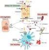
Regulation of systemic iron metabolism. Organs and cell types involved in systemic iron balance are shown. Duodenal enterocytes absorb dietary iron via DMT1 located on the apical surface upon reduction of Fe3+ to Fe2+ by DcytB. Spleenic reticuloendothelial macrophages recycle iron from senescent red blood cells. Both cell types release iron via ferroportin with the aid of hephaestin, which oxidizes Fe2+ to Fe3+. Iron is also oxidized by ceruloplasmin in the circulation. Plasma Tf captures and circulates iron in the body. Hepatic hormone, hepcidin regulates iron efflux from these cells by regulating the stability of ferroportin. Synthesis and secretion of hepcidin by hepatocytes is influenced by iron levels in the body as well as conditions that affect iron metabolism indirectly such as inflammation, ER stress, erythropoiesis, and hypoxia (see text for additional details).
Fig. 3
Fig. 3
Cellular iron metabolism. Most cells in the body obtain iron from circulating differic Tf. Iron-loaded holo-Tf binds to TfR1 on the cell surface and the complex undergoes endocytosis via clathrin-coated pits. A proton pump acidifies the endosome resulting in the release of Fe3+, which is subsequently reduced to Fe2+ by Steap3 and transported across the endosomal membrane to the cytosol by DMT1. DMT1 also facilitates dietary iron absorption. Apo-Tf is recycled back to the cell surface and released from TfR1 to plasma to repeat another cycle. Newly acquired iron enters into cytosolic “labile iron pool” (LIP), which is redox-active. LIP is chelated by intracellular siderophore that facilitates intracellular iron trafficking to mitochondria via an unknown receptor for metabolic utilization (such as synthesis of heme and iron-sulfur clusters), and cellular iron that is not utilized is either stored in ferritin or exported via ferroportin. Cells also export iron contained in ferritin and heme.
Fig. 4
Fig. 4
Cellular iron balance. A typical IRE motif consists of a hexanucleotide loop with the sequence 5′-CAGUGH-3′ (H could be A, C, or U) and a stem, interrupted by a bulge with an unpaired C residue. IREs post-transcriptional control expression of regulators of cellular iron metabolism in concert with IRPs. Translational-type IRE/IRP interactions in the 5′ UTR modulate the expression of the mRNAs encoding H- and L-ferritin, ALAS2, m-aconitase, ferroportin, and HIF-2α, which in turn control iron storage, erythroid iron utilization, energy homeostasis, iron efflux, and hypoxic responses, respectively. Conversely, IRE/IRP interactions in the 3′ UTR stabilize the mRNAs encoding TfR1, DMT1, and Cdc14A, which are involved in iron uptake, iron transport, and the cell cycle, respectively. Under physiological conditions, IRP1 is regulated by a reversible ISC switch. Iron deficiency, promotes ISC disassembly and a conformational rearrangement, resulting in conversion of IRP1 from c-aconitase to an IRE-binding protein. The ISC is regenerated in iron-replete cells. Hypoxia favors maintenance of the ISC, while H2O2 promotes its disassembly. When the ISC biogenesis pathway is not operational, iron leads to ubiquitination of apo-IRP1 by the FBXL5 E3 ligase complex (including Skp1, Cul1 and Rbx1), resulting in proteasomal degradation. IRP2 is stable in iron deficient and/or hypoxic cells; under these conditions FBXL5 undergoes ubiquitination and proteasomal degradation. An increase in iron and oxygen levels stabilizes FBXL5 by formation of an Fe-O-Fe center in its hemerythrin domain, triggering the assembly of an E3 ubiquitin ligase complex together with Skp1, Cul1 and Rbx1. This complex ubiquitinates IRP2, leading to its recognition by the proteasome and its degradation.

Similar articles

Cited by

References

    1. Theil EC, Raymond KN. Bioinorganic chemistry. In: Bertini I, Gray HB, Lippard SJ, Valentine JS, editors. University Science Books; Mill Valley, CA: 1994. pp. 1–36.
    1. Wigglesworth JM, Baum H. In: In Iron in Biochemistry and Medicine II. Jacobs A, Worwood M, editors. Academic press; New York, NY: 1980. pp. 29–86.
    1. Pereira IAC, Teixeira M, Xavier AV. In: Bioinorganic chemistry: Trace element evolution from anaerobes to aerobes. Williams RJP, editor. Springer-Verlag; New York, NY: 2010. pp. 91–190.
    1. Nielands JB. Siderophores: structure and functions of microbial iron transport compounds. J Biol Chem. 1995;27:26723–26726. - PubMed
    1. Hider RC, Kong XL. Chemistry and biology of siderophores. Nat Products Rep. 2010;27:637–657. - PubMed

Publication types