. 2012 Jun 15:13:31.
doi: 10.1186/1471-2172-13-31.

TGFβ signaling plays a critical role in promoting alternative macrophage activation

Affiliations

TGFβ signaling plays a critical role in promoting alternative macrophage activation

Dapeng Gong et al. BMC Immunol. .

Abstract

Background: Upon stimulation with different cytokines, macrophages can undergo classical or alternative activation to become M1 or M2 macrophages. Alternatively activated (or M2) macrophages are defined by their expression of specific gene products and play an important role in containing inflammation, removing apoptotic cells and repairing tissue damage. Whereas it is well-established that IL-4 can drive alternative activation, if lack of TGFβ signaling at physiological levels affects M2 polarization has not been addressed.

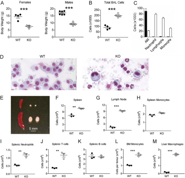
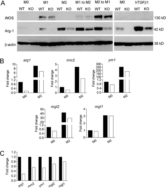
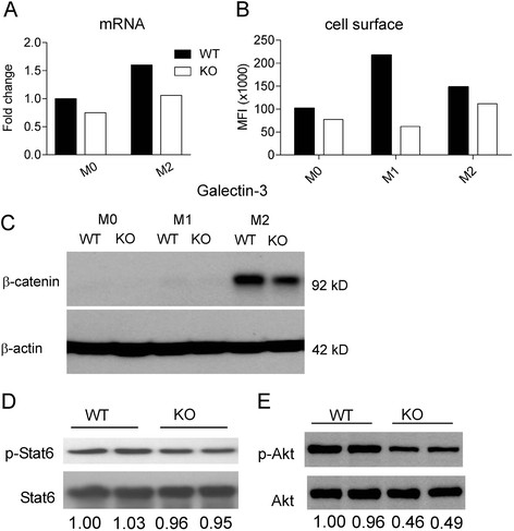
Results: Vav1-Cre x TβRIIfx/fx mice, lacking TβRII function in hematopoietic cells, exhibited uncontrolled pulmonary inflammation and developed a lethal autoimmune syndrome at young age. This was accompanied by significantly increased numbers of splenic neutrophils and T cells as well as elevated hepatic macrophage infiltration and bone marrow monocyte counts. TβRII-/- CD4+ and CD8+ T-cells in the lymph nodes and spleen expressed increased cell surface CD44, and CD69 was also higher on CD4+ lymph node T-cells. Loss of TβRII in bone marrow-derived macrophages (BMDMs) did not affect the ability of these cells to perform efferocytosis. However, these cells were defective in basal and IL-4-induced arg1 mRNA and Arginase-1 protein production. Moreover, the transcription of genes that are typically upregulated in M2-polarized macrophages, such as ym1, mcr2 and mgl2, was also decreased in peritoneal macrophages and IL-4-stimulated TβRII-/- BMDMs. We found that cell surface and mRNA expression of Galectin-3, which also regulates M2 macrophage polarization, was lower in TβRII-/- BMDMs. Very interestingly, the impaired ability of these null mutant BMDMs to differentiate into IL-4 polarized macrophages was Stat6- and Smad3-independent, but correlated with reduced levels of phospho-Akt and β-catenin.

Conclusions: Our results establish a novel biological role for TGFβ signaling in controlling expression of genes characteristic for alternatively activated macrophages. We speculate that lack of TβRII signaling reduces the anti-inflammatory M2 phenotype of macrophages because of reduced expression of these products. This would cause defects in the ability of the M2 macrophages to negatively regulate other immune cells such as T-cells in the lung, possibly explaining the systemic inflammation observed in Vav1-Cre x TβRIIfx/fx mice.


Figures

Figure 1
Figure 1
Mice lacking TβRII in hematopoietic cells develop a lethal inflammatory and autoimmune syndrome. (A) Body weight of male and female Vav-Cre x TβRIIfx/fx(TβRII-/-) mice and littermate controls at postnatal day 18-21. (B) Total cell counts and (C) differential cell counts in BALF of WT and TβRII-/- mice at postnatal day 18-21. Solid bar, WT; Open bar, TβRII-/- samples. Differences are significant (p < 0.01) for all cell populations in panel C. (D) Wright-Giemsa-stained BALF cells from the indicated genotypes (18-day old littermates). Note the presence of a single cell type - the alveolar macrophage - in the control lung, whereas the TβRII-/- sample contains neutrophils, monocytes and lymphocytes. (E) Photographic images of spleens and lymph nodes; total cell counts in (F) spleens and (G) lymph nodes; total numbers of (H) monocytes, (I) neutrophils, (J) T-cells and (K) B-cells in spleen; total counts of bone marrow monocytes (L) and liver macrophages (M) in WT (filled circles)and TβRII-/- (KO, open circles) mice (n = 3, **p < 0.01; ***p < 0.001).
Figure 2
Figure 2
Expression of M2 markers on BMDMs is impaired in the absence of TβRII signaling. (A) Western blot analysis of WT and TβRII-/- BMDMs cultured for 24 hrs with medium alone (M0), with LPS/IFNγ (M1), IL-4 (M2), or hTGFβ1. β-actin serves as a loading control. Increased Arg-1 in KO BMDM upon hTGFβ1stimulation due to residual WT cells, see Additional file 1: Figure S2E. (B) Real-time RT-PCR for the indicated genes after incubation for 24 hrs with medium alone (M0) or IL-4 (M2). (C) Real-time RT-PCR for the indicated genes in naïve peritoneal macrophages. Fold change is with respect to the expression level of M0 WT. Solid bar, WT; Open bar, TβRII-/- peritoneal macrophages. The results shown are representative of one of two independently derived sets of BMDMs from different mice.
Figure 3
Figure 3
Lack ofTβRIIsignaling is correlated with decreased Galectin-3, β-catenin and phospho-Akt levels. (A) Real-time RT-PCR for lgals3 in BMDMs. Fold change is with respect to the expression of M0 WT. Solid bar, WT; Open bar, TβRII-/-. (B) Cell surface expression of Galectin-3 on WT and TβRII-/- BMDMs measured using FACS. MFI, mean fluorescent intensity. The result shown is representative of one of two independently derived sets of BMDMs from different mice for both A and B. (C) Western blot analysis of β-catenin expression. BMDM were treated with medium alone (M0), LPS/IFNγ (M1), or IL-4 (M2). Results are representative of 3 pairs of matched WT and TβRII-/- mice. Western blot analysis of phosphorylated-Stat6 (D) and phosphorylated-Akt (E) in WT and TβRII-/- BMDMs that had been treated with IL-4 for 1 hr. Results are shown for 2 independent mice per genotype. The numbers at the bottom represent the band intensity ratio of phosphorylated protein versus total protein, normalized to lane 1 on each blot.

References

    1. Blobe GC, Schiemann WP, Lodish HF. Role of transforming growth factor beta in human disease. N Engl J Med. 2000;342:1350–1358. doi: 10.1056/NEJM200005043421807. - DOI - PubMed
    1. Iseki S, Osumi-Yamashita N, Miyazono K, Franzen P, Ichijo H, Ohtani H, Hayashi Y, Eto K. Localization of transforming growth factor-beta type I and type II receptors in mouse development. Exp Cell Res. 1995;219:339–347. doi: 10.1006/excr.1995.1237. - DOI - PubMed
    1. Millan FA, Denhez F, Kondaiah P, Akhurst RJ. Embryonic gene expression patterns of TGF beta 1, beta 2 and beta 3 suggest different developmental functions in vivo. Development. 1991;111:131–143. - PubMed
    1. Roberts AB, Sporn MB. Differential expression of the TGF-beta isoforms in embryogenesis suggests specific roles in developing and adult tissues. Mol Reprod Dev. 1992;32:91–98. doi: 10.1002/mrd.1080320203. - DOI - PubMed
    1. Li MO, Wan YY, Sanjabi S, Robertson AK, Flavell RA. Transforming growth factor-beta regulation of immune responses. Annu Rev Immunol. 2006;24:99–146. doi: 10.1146/annurev.immunol.24.021605.090737. - DOI - PubMed

Publication types

MeSH terms

Substances