. 2012 Jul 4;134(26):10717-20.
doi: 10.1021/ja300624j. Epub 2012 Jun 22.

Evidence for helical structure in a tetramer of α2-8 sialic acid: unveiling a structural antigen

Affiliations

Evidence for helical structure in a tetramer of α2-8 sialic acid: unveiling a structural antigen

Marcos D Battistel et al. J Am Chem Soc. .

Abstract

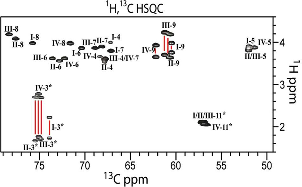
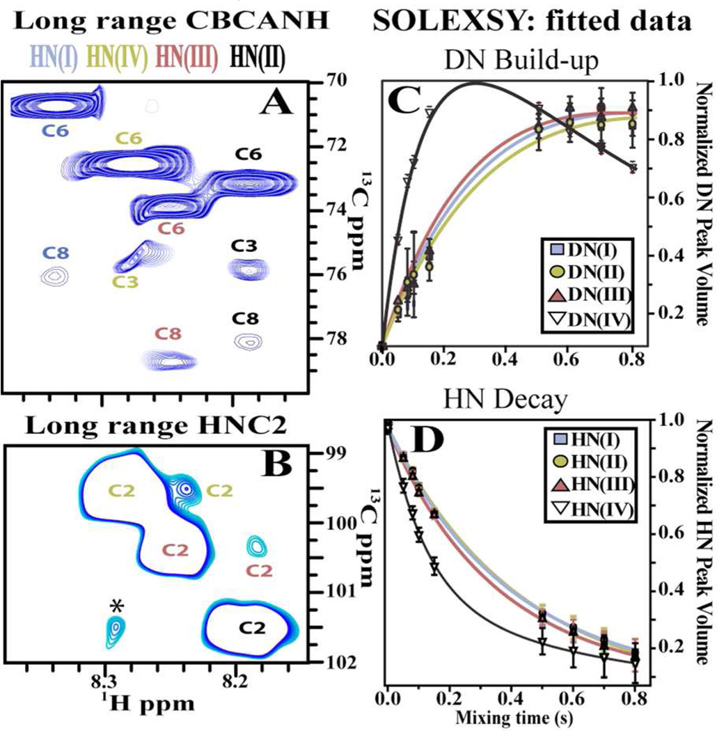
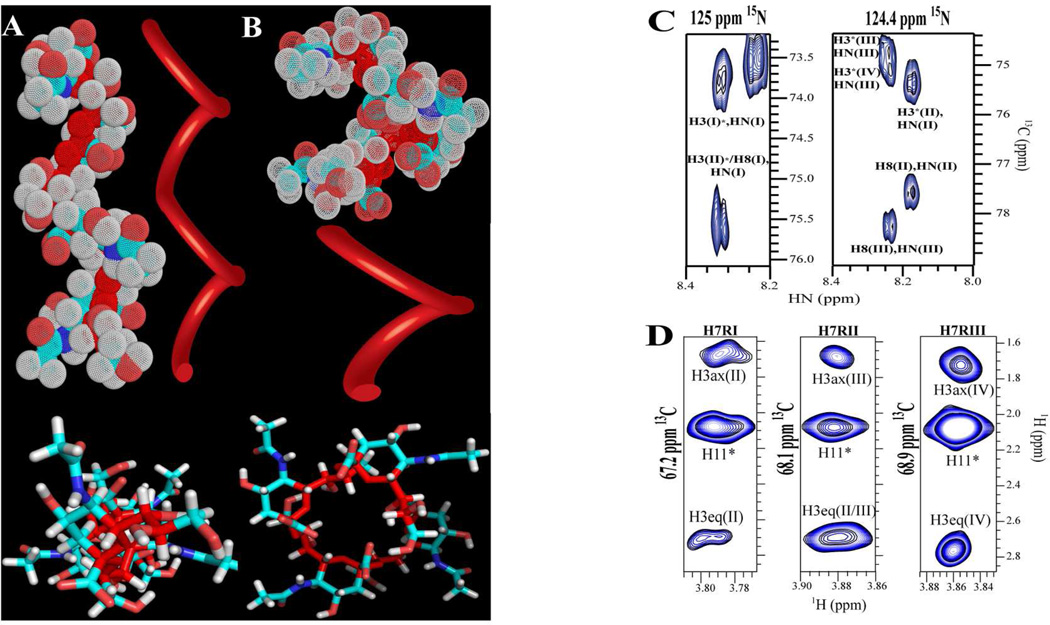
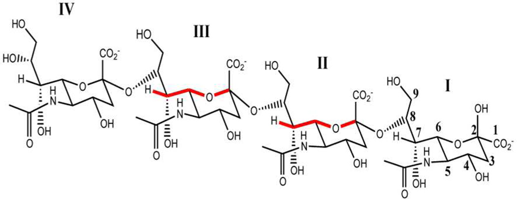
Characteristic H-bonding patterns define secondary structure in proteins and nucleic acids. We show that similar patterns apply for α2-8 sialic acid (SiA) in H(2)O and that H-bonds define its structure. A (15)N,(13)C α2-8 SiA tetramer, (SiA)(4), was used as a model system for the polymer. At 263 K, we detected intra-residue through-H-bond J couplings between (15)N and C8 for residues R-I-R-III of the tetramer, indicating H-bonds between the (15)N's and the O8's of these residues. Additional J couplings between the (15)N's and C2's of the adjacent residues confirm the putative H-bonds. NH groups showing this long-range correlation also experience slower (1)H/(2)H exchange. Additionally, detection of couplings between H7 and C2 for R-II and R-III implies that the conformations of the linkers between these residues are different than in the monomers. These structural elements are consistent with two left-handed helical models: 2 residues/turn (2(4) helix) and 4 residues/turn (1(4) helix). To discriminate between models, we resorted to (1)H,(1)H NOEs. The 2(4) helical model is in better agreement with the experimental data. We provide direct evidence of H-bonding for (SiA)(4) and show how H-bonds can be a determining factor for shaping its 3D structure.


Figures

Figure 1
Figure 1
1H,13C-HSQC spectrum of (SiA)4. The spectrum, collected at 263K and pH 6.5 (20 mM Na2HPO4), displays excellent dispersion and signal-to-noise. Assignments are labeled with Roman numerals, to indicate residue position, followed by Arabic numerals to indicate atom position.
Figure 2
Figure 2
NMR experiments for hydrogen bond detection (263K pH* 6.5). A) Long range CBCANH: Correlations to different (SiA)4 residues are color coded based on residue position. B) Long range HNC2 on (SiA)4. Asterisk denotes an impurity. SOLEXSY DN build-up (C) and HN decay (D) traces obtained for each (SiA)4 residue. The data were fitted to extract exchange rate constants presented on Table 1.
Figure 3
Figure 3
Panels A and B show two different static models for (SiA)4 consistent with the experimental data. Top figures depict side views of helical models with two (Panel A) and 4 residues per turn (Panel B). Bottom figures show a top view of the models in stick representation. Oligosaccharide backbone is depicted in dark red. A ribbon was placed proximal to each model to depict the trace followed by the oligosaccharide backbone. C) CNH-NOESY spectrum: though-space intra and inter-residue correlations observed between amide hydrogen and ring hydrogen atoms. D) 1H,13C-HSQC-NOESY: NOE correlations between H7 of each of the first three residues to H3s of a contiguous residue.
Chart 1
Chart 1
α2-8 Sialic acid tetramer (SiA)4 chemical structure. W-coupling is highlighted in red for residues for which it was detected.

Similar articles

Cited by

References

    1. Hill DJ, Griffiths NJ, Borodina E, Virji M. Clin. Sci. 2010;118:547. - PMC - PubMed
    1. Echenique-Rivera H, Muzzi A, Tordello ED, Seib KL, Francois P, Rappuoli R, Pizza M, Serruto D. PLOS Pathogens. 2011;7 e10020271. - PMC - PubMed
    1. Kim KJ, Elliott SJ, Cello FD, Stins MF, Kim KS. Cell Microbiol. 2003;5:245. - PubMed
    1. Wyle FA, Tramont EC, Lowentha Jp, Berman SL, Brandt BL, Altieri PL, Artenste Ms, Kasper DL. J Infect Dis. 1972;126:514. - PubMed
    1. Finne J, Leinonen M, Makela PH. Lancet. 1983;2:355. - PubMed

MeSH terms