

doi: 10.1016/j.stem.2012.05.012.
Adult neural stem cells bridge their niche
Affiliations
- PMID: 22704510
- PMCID: PMC3726005
- DOI: 10.1016/j.stem.2012.05.012
Item in Clipboard
Adult neural stem cells bridge their niche
Cell Stem Cell.
.
Abstract
Major developments in the neural stem cell (NSC) field in recent years provide new insights into the nature of the NSC niche. In this perspective, we integrate recent anatomical data on the organization of the two main neurogenic niches in the adult brain, the ventricular-subventricular zone (V-SVZ) and the subgranular zone (SGZ), with signaling pathways that control the behavior of NSCs. NSCs in the adult brain stretch into physiologically distinct compartments of their niche. We propose how adult NSCs' morphology may allow these cells to integrate multiple signaling pathways arising from unique locations of their niche.
Copyright © 2012 Elsevier Inc. All rights reserved.
Figures
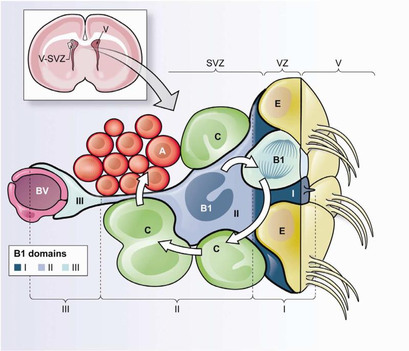
Upper left. Frontal cross-section of the adult mouse brain showing the location of the ventricularsubventricular zone (V-SVZ), where neurogenesis in walls of the lateral ventricles (V) continues throughout life. Lower panel. Cellular composition of the adult V-SVZ niche and domains of B1 cells. Neural stem cells (NSCs) correspond to type B1 cells (blue). B1 cells are surrounded by multiciliated ependymal cells (E) forming pinwheel-like structures on the ventricular surface. B1 cells give rise to intermediate progenitors (IPCs or C cells, green), which correspond to transit-amplifying cells that divide to generate neuroblasts (Type A cells, red). B1 cells retain epithelial properties, with a thin apical process (containing a primary cilium) that contacts the lateral ventricle (V), and a long basal process ending on blood vessels (BV, purple). Therefore, B1 cells can be subdivided into three domains. Domain I (proximal or apical, dark blue) contains the primary cilium and is in direct contact with the CSF; within this domain B1 cells can access soluble factors within the CSF and signaling molecules from neighboring ependymal cells. Domain II (intermediate, medium blue) is in close proximity to IPCs, neuroblasts, neuronal terminals and other supporting cells; cell-cell interactions between B1 cells and their progeny could occur within this compartment. Domain III (distal, light blue) comprises a basal process ending in a specialized end-foot that contacts blood vessels; blood-borne factors and endothelial-derived factors may act on B1 cells in this domain.

Upper panel. Frontal cross-section of the adult mouse brain showing the hippocampal formation (left). The insert shows a higher magnification indicating the location of the dentate gyrus (right). Lower panel. Cellular composition of the adult dentate gyrus and domains of SGZ radial astrocytes. Radial astrocytes (RA, also known as type 1 cells (blue)) give rise to intermediate progenitors (IPCs, green), which progressively (via IPC1 and IPC2 (type 2a and type 2b cells)) differentiate into immature granule cells (IGCs (type 3 cells), red). Mature granule cells (GCs, brown) send an axon parallel to the SGZ into the hilus, while their dendrites branch into the ML. Radial astrocytes are polarized cells with their cell body in the SGZ, a long main shaft that traverses through the granule cell layer (GCL) and then branches diffusely in the inner molecular layer (IML). Here we subdivide radial astrocytes into three domains. Domain I (proximal, dark blue) faces the hilus, harbors a primary cilium, contacts with blood vessels (BV, purple) and through lateral processes, neighboring radial astrocytes. Factors derived from blood, endothelial cells and neighboring radial astrocytes act on NSCs within this domain. Domain II (intermediate, medium blue) contains the cell body and the main shaft. This part of the cell interacts closely with IPCs and GCs; this domain allow specific cell-cell interactions of NSCs with their progeny and detection of local neural activity and signaling from GCs. Domain III (distal, light blue) contacts other glial cells, axons, and synaptic terminals in the inner molecular layer; NSCs may detect levels of neural activity from Mossy cells and other neurons within this compartment.
References
-
- Alfonso J, Le Magueresse C, Zuccotti A, Khodosevich K, Monyer H. Diazepam binding inhibitor promotes progenitor proliferation in the postnatal SVZ by reducing GABA signaling. Cell Stem Cell. 2012;10:76–87. - PubMed
-
- Altman J. The Discovery of Adult Mammalian Neurogenesis. In: Seki T, Sawamoto K, Parent JM, Alvarez-Buylla A, editors. Neurogenesis in the Adult Brain I: Neurobiology. Springer; Tokyo: 2011. pp. 3–46.
Publication types
MeSH terms
Grants and funding
LinkOut - more resources
Full Text Sources
Other Literature Sources
