. 2012 Jun 14;11(6):664-73.
doi: 10.1016/j.chom.2012.04.018.

Monitoring the inflammatory response to infection through the integration of MALDI IMS and MRI

Affiliations

Monitoring the inflammatory response to infection through the integration of MALDI IMS and MRI

Ahmed S Attia et al. Cell Host Microbe. .

Abstract

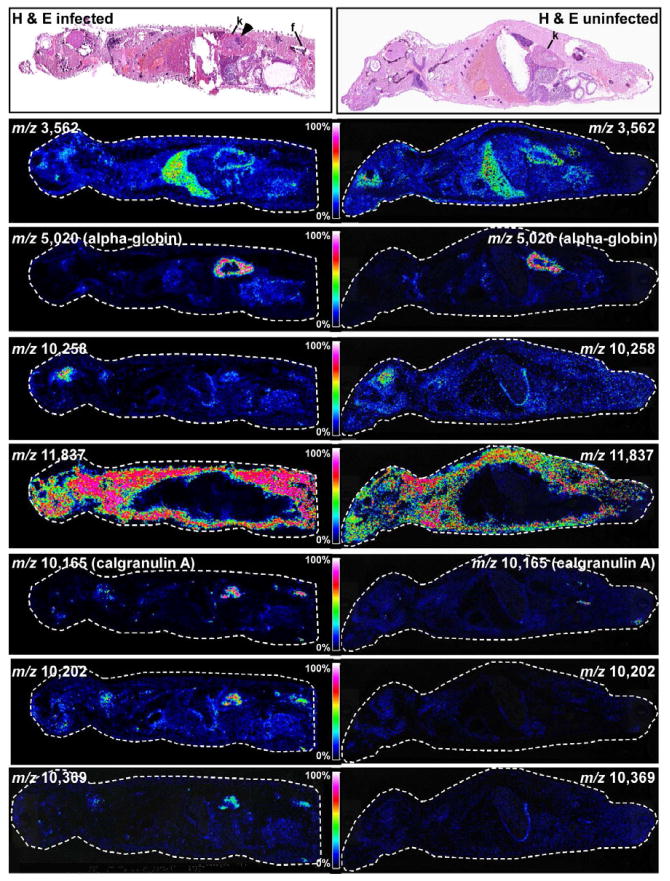
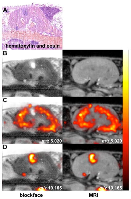
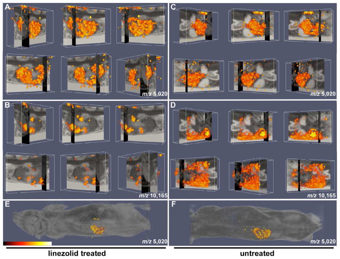
Systemic bacterial infection is characterized by a robust whole-organism inflammatory response. Analysis of the immune response to infection involves technologies that typically focus on single organ systems and lack spatial information. Additionally, the analysis of individual inflammatory proteins requires antibodies specific to the protein of interest, limiting the panel of proteins that can be analyzed. Herein we describe the application of matrix-assisted laser desorption/ionization imaging mass spectrometry (MALDI IMS) to mice systemically infected with Staphylococcus aureus to identify inflammatory protein masses that respond to infection throughout an entire infected animal. Integrating the resolution afforded by magnetic resonance imaging (MRI) with the sensitivity of MALDI IMS provides three-dimensional spatially resolved information regarding the distribution of innate immune proteins during systemic infection, allowing comparisons to in vivo structural information and soft-tissue contrast via MRI. Thus, integrating MALDI IMS with MRI provides a systems-biology approach to study inflammation during infection.


Conflict of interest statement

The authors have no conflicts of interest to report.

Figures

Figure 1
Figure 1. Linezolid reduces staphylococcal abscess formation and inflammation in systemically infected mice
Groups of three animals were infected with S. aureus for 96 hours at which point they were either treated with 0.2 mg/ml linezolid or left untreated and the infection was allowed to proceed for an additional 96 hours. (A) Bacterial burdens in the kidneys of the infected mice were estimated by tissue homogenization and plating. (B and C) The percentage of neutrophils (B) and macrophages (C) in the kidneys of the two groups of mice as determined by fluorescently activated cell sorting. Error bars in panels A–C represent +/− standard deviation. (D) Photographic image of representative kidneys from the treated and control mice. (E and F) Hematoxylin and eosin stain of kidney sections from control (E) and treated (F) mice 8 days following infection. Black arrows denote abscesses.
Figure 2
Figure 2. MRI of S. aureus infected mice that have been treated with linezolid or left untreated
Four contiguous slices depicting the kidneys in the coronal plane for one treated and one untreated animal, acquired using a 3D gradient echo imaging sequence at 9.4T in approximately 33 minutes. Panels (A) and (B) show one infected animal on day 4 (just prior to treatment with linezolid) and day 8, respectively. Abscesses are marked with arrows. Panels (C) and (D) show an untreated animal on day 4 and day 8, respectively.
Figure 3
Figure 3. Whole animal MALDI IMS of the systemic response to S. aureus infection
(Top panels) Hematoxylin and eosin stained sections of entire mice that were either infected with S. aureus or left uninfected. Abscesses are marked by an arrow. Kidneys are marked with “k” and femur is marked with “f”. Masses corresponding to proteins that are abundant in the liver (m/z 3,562), kidney (m/z 5,020), brain (m/z 10,258), or systemically (m/z 11,837) in both infected and uninfected mice are shown. In addition, masses corresponding to proteins that are only expressed in infected animals are shown (m/z 10,165, 10,202, 10,369). The color scale correlates to 0–100% full scale on the intensity of each ion.
Figure 4
Figure 4. Co-registered MALDI IMS and MRI reveals proteins that exhibit disease state distribution patterns
(A) Hematoxylin and eosin stained section showing the presence of kidney abscesses. (B–D) Blockface image (left) and MRI (right) co-registered with MALDI IMS signals at (C) m/z 5,020 (alpha-globin), or (D) m/z 10,165 (calgranulin A). The data are presented as arbitrary units of intensity from 0 (dark red) to 1 (white).
Figure 5
Figure 5. Three-dimensional integration of MALDI IMS and MRI for imaging the inflammatory response to infection
(A–B) Orthogonal blockface and MRI slice data of linezolid-treated mouse with overlaid (A) alpha-globin protein density (m/z 5,020) and (B) calgranulin A protein density (m/z 10,165) volume renderings. (C–D) Orthogonal blockface and MRI slice data of untreated mouse with overlaid (C) alpha-globin protein density (m/z 5,020) and (D) calgranulin A protein density (m/z 10,165) volume renderings. (E and F) Protein density (m/z 5,020) from (E) linezolid-treated and (F) untreated mice superimposed on whole mouse image. The data in all panels are presented as arbitrary units of intensity from 0 (dark red) to 1 (white). See also Supplemental Movies S1–4.

Comment in

  • Inflammation in 3D.
    Kobayashi SD, DeLeo FR. Kobayashi SD, et al. Cell Host Microbe. 2012 Jun 14;11(6):557-9. doi: 10.1016/j.chom.2012.05.006. Cell Host Microbe. 2012. PMID: 22704615

Similar articles

Cited by

References

    1. Andersson M, Groseclose MR, Deutch AY, Caprioli RM. Imaging mass spectrometry of proteins and peptides: 3D volume reconstruction. Nature Methods. 2008;5:101–108. - PubMed
    1. Bernardo K, Pakulat N, Fleer S, Schnaith A, Utermohlen O, Krut O, Muller S, Kronke M. Subinhibitory concentrations of linezolid reduce Staphylococcus aureus virulence factor expression. Antimicrob Agents Chemother. 2004;48:546–555. - PMC - PubMed
    1. Cheng AG, Kim HK, Burts ML, Krausz T, Schneewind O, Missiakas DM. Genetic requirements for Staphylococcus aureus abscess formation and persistence in host tissues. FASEB J. 2009;23:3393–3404. - PMC - PubMed
    1. Corbin BD, Seeley EH, Raab A, Feldmann J, Miller MR, Torres VJ, Anderson KL, Dattilo BM, Dunman PM, Gerads R, et al. Metal Chelation and Inhibition of Bacterial Growth in Tissue Abscesses. Science. 2008;319:962–965. - PubMed
    1. DeLeo FR, Otto M, Kreiswirth BN, Chambers HF. Community-associated meticillin-resistant Staphylococcus aureus. The Lancet. 2010;375:1557–1568. - PMC - PubMed

Publication types

MeSH terms