Review
. 2012 Aug;143(2):307-20.
doi: 10.1053/j.gastro.2012.06.004. Epub 2012 Jun 13.

A liver full of JNK: signaling in regulation of cell function and disease pathogenesis, and clinical approaches

Affiliations
Review

A liver full of JNK: signaling in regulation of cell function and disease pathogenesis, and clinical approaches

Ekihiro Seki et al. Gastroenterology. 2012 Aug.

Abstract

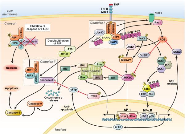
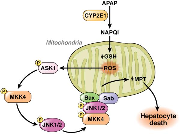
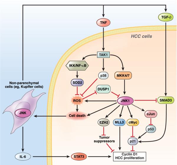
c-Jun-N-terminal kinase (JNK) is a mitogen-activated protein kinase family member that is activated by diverse stimuli, including cytokines (such as tumor necrosis factor and interleukin-1), reactive oxygen species (ROS), pathogens, toxins, drugs, endoplasmic reticulum stress, free fatty acids, and metabolic changes. Upon activation, JNK induces multiple biologic events through the transcription factor activator protein-1 and transcription-independent control of effector molecules. JNK isozymes regulate cell death and survival, differentiation, proliferation, ROS accumulation, metabolism, insulin signaling, and carcinogenesis in the liver. The biologic functions of JNK are isoform, cell type, and context dependent. Recent studies using genetically engineered mice showed that loss or hyperactivation of the JNK pathway contributes to the development of inflammation, fibrosis, cancer growth, and metabolic diseases that include obesity, hepatic steatosis, and insulin resistance. We review the functions and pathways of JNK in liver physiology and pathology and discuss findings from preclinical studies with JNK inhibitors.


Figures

Figure 1
Figure 1. The JNK Activation Pathway and Domain Structure and
A. number of stimuli, including growth factors, cytokines, PAMPs, ROS, and environmental stresses activate the JNK signaling pathway through a 3-tier kinase cascade that includes MAP3Ks (ASK1, TAK1, MEKKs and MLKs), MAP2Ks (MKK4 and MKK7), and JNKs. Activated JNKs phosphorylate their substrates, which include transcription factors (c-Jun, JunB, JunD, p53, and c-Myc) and mitochondrial proteins (Bcl-2, Bid, Bim, Bax, and Mcl-1), to induce various biological responses. B. The JNK protein contains a conserved dual phosphorylation Thr-Pro-Tyr motif in its activation loop and has a common substrate docking site in C-terminus and a glutamate/aspartate domain in its N-terminus to mediate interactions with MAP2Ks, phosphatases, and substrates.
Figure 2
Figure 2. JNKs in TNF Signaling
(1) Binding of TNF to the TNFR type I leads to the rapid formation of complex I, comprising TRADD, RIP1, TRAF2, cIAP1, cIAP2, and Ubc13. cIAP-mediated K63-ubiquitination of RIP1 recruits and activates TAK1. (2) MAP3Ks (TAK1 and ASK1) activate JNK1 and 2 through MKK4 and 7. JNK activates AP-1, which comprises c-Jun and c-Fos. Simultaneously, JNK1 phosphorylates ITCH to ubiquitinate c-FLIP, which promotes caspase-8 dependent apoptosis. JNKs can also induce mitochondria-dependent apoptosis through Bax and degradation of Bim. (3) TAK1 phosphorylates and activates the IKK complex, which leads to phosphorylation, unbiquitination, and degradation of IκBα, resulting in nuclear translocation and activation of NF-κB, which is comprises the p50 and p65 subunits. NF-κB induces the transcription of SOD2 and c-FLIP to prevent ROS production and caspase-8 activation, respectively. (4) Complex I also contributes to ROS production through NOX1 and Rac1. (5) Following formation of complex I, RIP1 is deubiquitinated by CYLD or A20 to form complex II, comprising TRADD, FADD, RIP1, RIP3, and caspase-8. Normally, caspase-8 induces apoptosis. (6) However, if caspase-8 or FADD is blocked, RIP1 and RIP3 are phosphorylated and cause necrosis.
Figure 3
Figure 3. Role of JNK in Acetaminophen-Induced Liver Injury
Acetaminophen is metabolized to NAPQI through CYP2E1, which reduces glutathione levels in mitochondria. Excessive NAPQI induces ROS, which activates ASK1, MKK4, and JNK. JNK, MKK4 and Bax translocate to the outer membrane of mitochondria through binding of Sab to increase the mitochondrial permeability transition, resulting in induction of massive hepatocyte death.
Figure 4
Figure 4. Roles of JNKs in the Pathogenesis of NAFLD
Obesity and hyperlipidemia increase plasma levels of FFA, which activate MLK3 and JNK in hepatocytes. JNK1 contributes to hepatic insulin resistance by phosphorylating IRS-1 at serine 307, and mitochondria-mediated hepatocyte death through Bax and PUMA. Saturated FFAs activate JNK through oxidative stress, ER stress, and lipid peroxidation in hepatocytes. Saturated FFAs also activate JNK in inflammatory cells to contribute to production of inflammatory cytokines. TLR4 on inflammatory cells might be involved in FFA-induced JNK activation.
Figure 5
Figure 5. Functions of JNKs in HCC
In HCC, JNK1 is overactivated either by overactivation of the upstream kinases MKK4 and 7 or the inactivation of DUSP1. Activated JNK1 induces c-Myc, which suppresses p21 expression and promotes HCC proliferation via cyclin D1 expression. JNK1 also upregulates MLL3 and EZH2, 2 histone H3 methyl transferases to increase expression of the cell cycle-associated genes, such as cyclins, and inhibit the transcription of tumor suppressors, respectively. c-Jun inhibits p53 activity post-transcriptionally. JNK1 increase generation of ROS, which is inhibited by p38α and IKKβ activation of NF-κB, through induction of SOD2. JNK1 also inhibits TGF-β–induced Smad3 activation, thereby inducing p21 to suppress HCC promotion. All of these pathways regulate hepatocyte death, which results in the release of IL-6, TNF, and TGF-β through JNK1 in non-parenchymal cells, including Kupffer cells. IL-6 induces HCC proliferation via activation of STAT3in hepatocytes. JNK1 promotes (but TAK1, p38α, and IKKβ activation of NF-κB prevent) hepatocyte death and HCC formation.

References

    1. Wagner EF, Nebreda AR. Signal integration by JNK and p38 MAPK pathways in cancer development. Nat Rev Cancer. 2009;9:537–49. - PubMed
    1. Davis RJ. Signal transduction by the JNK group of MAP kinases. Cell. 2000;103:239–52. - PubMed
    1. Kallunki T, Deng T, Hibi M, Karin M. c-Jun can recruit JNK to phosphorylate dimerization partners via specific docking interactions. Cell. 1996;87:929–39. - PubMed
    1. Haeusgen W, Herdegen T, Waetzig V. The bottleneck of JNK signaling: molecular and functional characteristics of MKK4 and MKK7. Eur J Cell Biol. 2011;90:536–44. - PubMed
    1. Bogoyevitch MA, Kobe B. Uses for JNK: the many and varied substrates of the c-Jun N-terminal kinases. Microbiol Mol Biol Rev. 2006;70:1061–95. - PMC - PubMed

Publication types

MeSH terms