

NPY and NPY receptors in airway structural and inflammatory cells in allergic asthma
- PMID: 22705097
- PMCID: PMC3488603
- DOI: 10.1016/j.yexmp.2012.05.009
NPY and NPY receptors in airway structural and inflammatory cells in allergic asthma
Abstract
Purpose: Neuropeptide Y (NPY) level is elevated in allergic asthmatic airways and activation of NPY receptor-1 (NPY-Y1) on antigen-presenting cells (APCs) is essential for T cell priming. Paradoxically, NPY-Y1 modulates hyper-responsiveness in T cells, suggesting a bimodal role for NPY in APCs and T cells. Therefore, determination of the temporal and spatial expression pattern of NPY and its receptors in asthmatic airways is essential to further understand the role of NPY in allergic asthma.
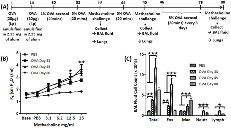
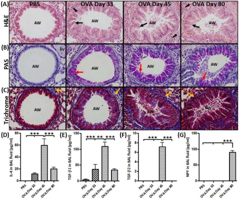
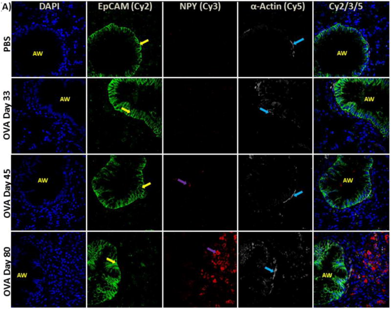
Methods: Lungs were isolated from control and acute and chronic stages of OVA-sensitized and challenged mice (OVA). Stains, including H&E, PAS, and trichrome, were used to determine the severity of lung pathology. The expression patterns of NPY and NPY-Y receptors in the airways were determined using ELISA and immunofluorescence. Cytokine levels in the BALF were also measured.
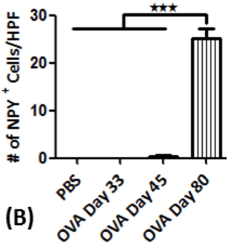
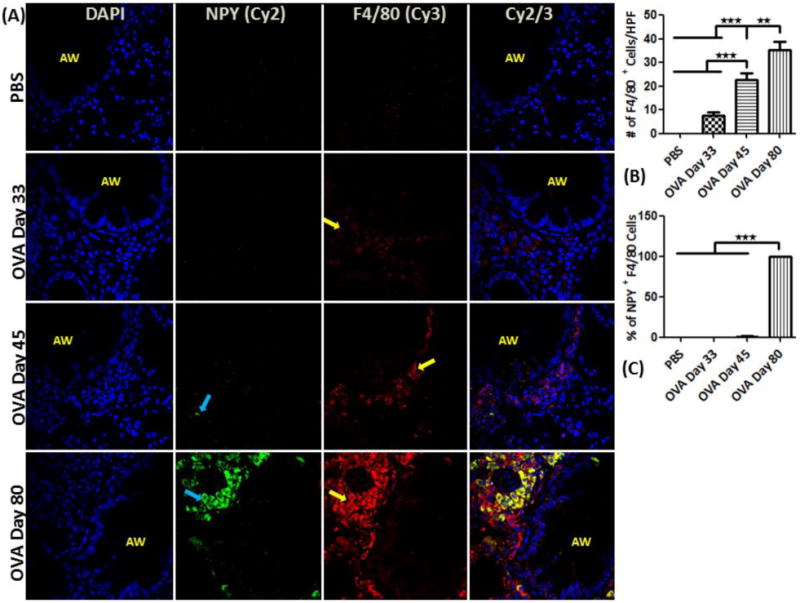
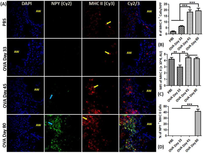
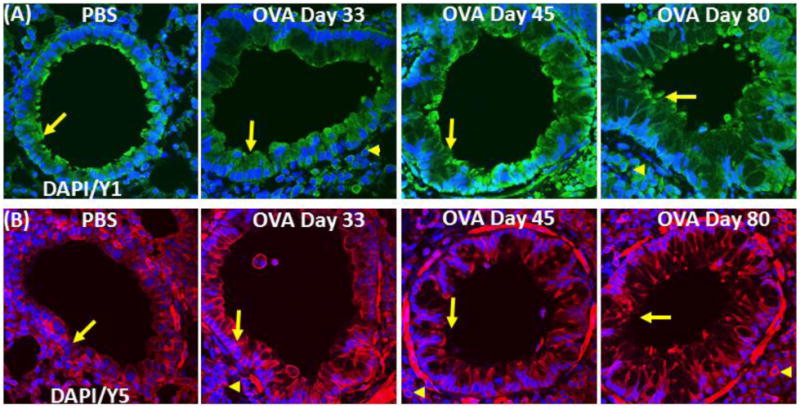
Results: NPY levels were undetectable in the BALF of control mice, but significantly increased in the OVA group at day 80. Levels of IL-4, TGF-β1 and TGF-β2, significantly increased and peaked on day 45 and decreased on day 80 in the OVA group, exhibiting an inverse correlation with NPY levels. NPY expression was localized to macrophage-like cells in the peri-bronchial and peri-vascular areas in the lung tissue. NPY-Y1 and -Y5 receptors were constitutively expressed by both structural and inflammatory cells in the lung tissue.
Conclusions: NPY produced by activated macrophage-like cells may be involved in regulating cytokine production and cellular activities of immune cells in asthma. However, it remains unclear whether such an increase in NPY is a defensive/compensatory mechanism to modulate the effects of inflammatory cytokines.
Copyright © 2012 Elsevier Inc. All rights reserved.
Figures
Similar articles
-
Regulatory Peptides in Asthma.Int J Mol Sci. 2021 Dec 20;22(24):13656. doi: 10.3390/ijms222413656. Int J Mol Sci. 2021. PMID: 34948451 Free PMC article. Review.
-
Y1 signalling has a critical role in allergic airway inflammation.Immunol Cell Biol. 2011 Nov;89(8):882-8. doi: 10.1038/icb.2011.6. Epub 2011 Mar 8. Immunol Cell Biol. 2011. PMID: 21383768
-
Requirement for neuropeptide Y in the development of type 2 responses and allergen-induced airway hyperresponsiveness and inflammation.Am J Physiol Lung Cell Mol Physiol. 2019 Mar 1;316(3):L407-L417. doi: 10.1152/ajplung.00386.2018. Epub 2019 Jan 3. Am J Physiol Lung Cell Mol Physiol. 2019. PMID: 30604629
-
Experimental protocol for development of adjuvant-free murine chronic model of allergic asthma.J Immunol Methods. 2019 May;468:10-19. doi: 10.1016/j.jim.2019.03.002. Epub 2019 Mar 14. J Immunol Methods. 2019. PMID: 30880263
-
The Roles of Neuropeptide Y in Respiratory Disease Pathogenesis via the Airway Immune Response.Acta Med Okayama. 2024 Apr;78(2):95-106. doi: 10.18926/AMO/66912. Acta Med Okayama. 2024. PMID: 38688827 Review.
Cited by
-
Clinical view on the importance of dendritic cells in asthma.Expert Rev Clin Immunol. 2013 Oct;9(10):899-919. doi: 10.1586/1744666X.2013.837260. Expert Rev Clin Immunol. 2013. PMID: 24128155 Free PMC article. Review.
-
Glucocorticoid Insensitivity in Severe Asthma: Underlying Molecular Mechanisms, Challenges, and Emerging Therapies.Arch Intern Med Res. 2025;8(2):107-120. doi: 10.26502/aimr.0202. Epub 2025 Apr 11. Arch Intern Med Res. 2025. PMID: 40337626 Free PMC article.
-
Multiple Regulatory Signals and Components in the Modulation of Bicarbonate Transporters.Pharmaceutics. 2024 Jan 5;16(1):78. doi: 10.3390/pharmaceutics16010078. Pharmaceutics. 2024. PMID: 38258089 Free PMC article. Review.
-
Sympathetic System in Wound Healing: Multistage Control in Normal and Diabetic Skin.Int J Mol Sci. 2023 Jan 20;24(3):2045. doi: 10.3390/ijms24032045. Int J Mol Sci. 2023. PMID: 36768369 Free PMC article. Review.
-
Regulatory Peptides in Asthma.Int J Mol Sci. 2021 Dec 20;22(24):13656. doi: 10.3390/ijms222413656. Int J Mol Sci. 2021. PMID: 34948451 Free PMC article. Review.
References
-
- Bedoui S, Miyake S, Straub RH, von Horsten S, Yamamura T. More sympathy for autoimmunity with neuropeptide Y? Trends in immunology. 2004;25:508–512. - PubMed
-
- El Karim IA, Linden GJ, Orr DF, Lundy FT. Antimicrobial activity of neuropeptides against a range of micro-organisms from skin, oral, respiratory and gastrointestinal tract sites. Journal of neuroimmunology. 2008;200:11–16. - PubMed
-
- Ericsson A, Hemsen A, Lundberg JM, Persson H. Detection of neuropeptide Y-like immunoreactivity and messenger RNA in rat platelets: the effects of vinblastine, reserpine, and dexamethasone on NPY expression in blood cells. Experimental cell research. 1991;192:604–611. - PubMed
-
- Jacob HJ, Pettersson A, Wilson D, Mao Y, Lernmark A, Lander ES. Genetic dissection of autoimmune type I diabetes in the BB rat. Nature genetics. 1992;2:56–60. - PubMed
Publication types
MeSH terms
Substances
Grants and funding
LinkOut - more resources
Full Text Sources
Other Literature Sources
Medical
Research Materials
Miscellaneous
