. 2012 Jun 29;36(6):959-73.
doi: 10.1016/j.immuni.2012.03.022. Epub 2012 Jun 14.

Ubiquitin-induced oligomerization of the RNA sensors RIG-I and MDA5 activates antiviral innate immune response

Affiliations

Ubiquitin-induced oligomerization of the RNA sensors RIG-I and MDA5 activates antiviral innate immune response

Xiaomo Jiang et al. Immunity. .

Abstract

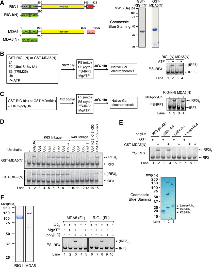
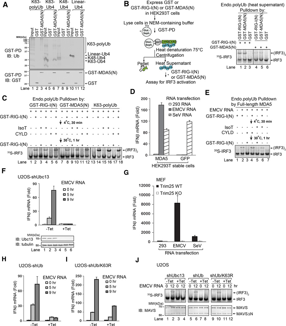
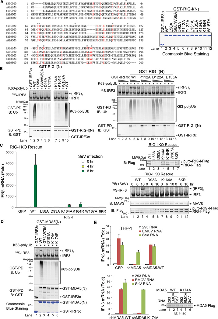
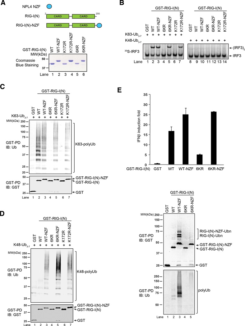
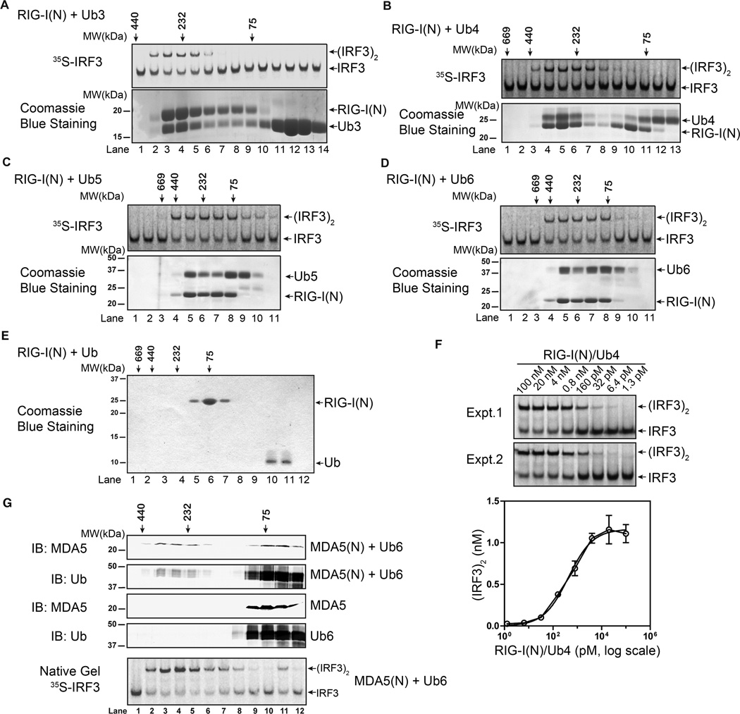
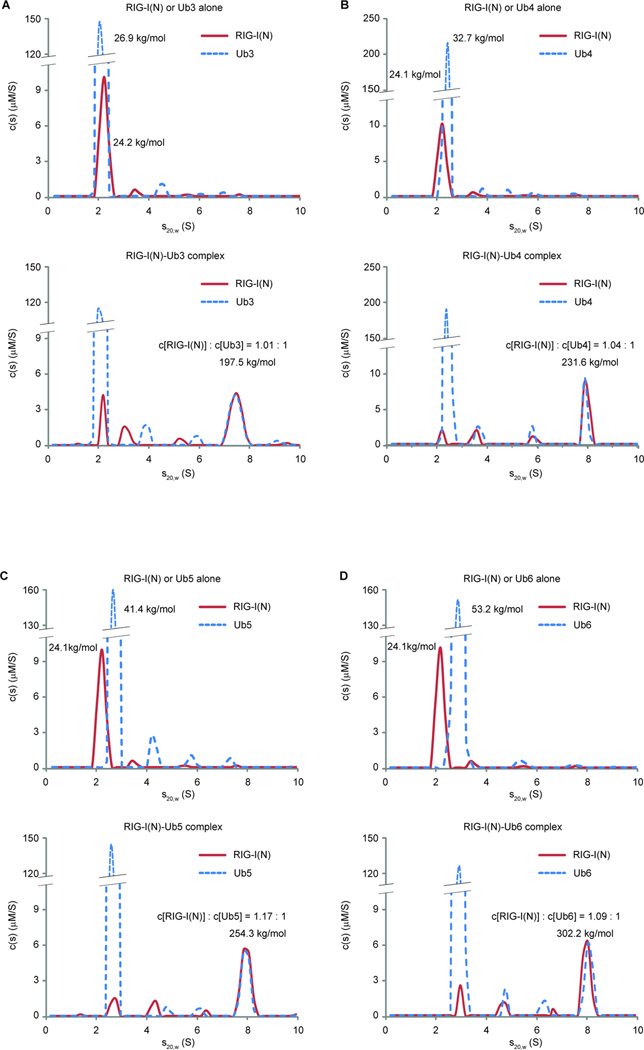
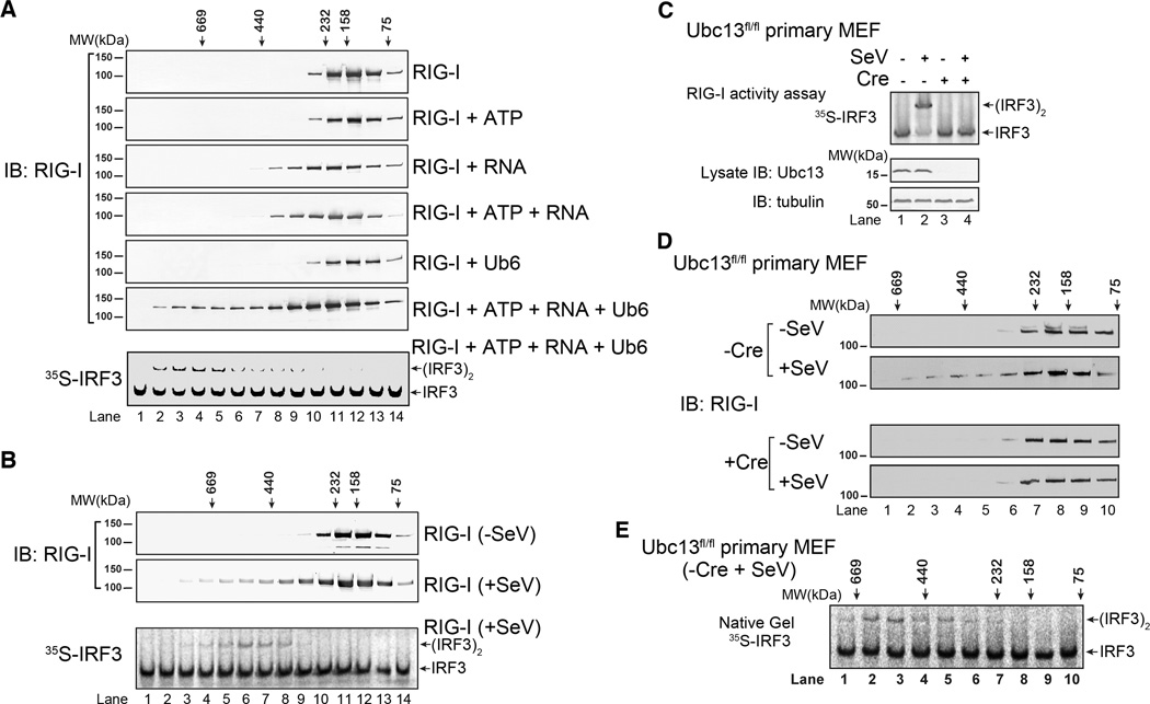
RIG-I and MDA5 detect viral RNA in the cytoplasm and activate signaling cascades leading to the production of type-I interferons. RIG-I is activated through sequential binding of viral RNA and unanchored lysine-63 (K63) polyubiquitin chains, but how polyubiquitin activates RIG-I and whether MDA5 is activated through a similar mechanism remain unresolved. Here, we showed that the CARD domains of MDA5 bound to K63 polyubiquitin and that this binding was essential for MDA5 to activate the transcription factor IRF3. Mutations of conserved residues in MDA5 and RIG-I that disrupt their ubiquitin binding also abrogated their ability to activate IRF3. Polyubiquitin binding induced the formation of a large complex consisting of four RIG-I and four ubiquitin chains. This hetero-tetrameric complex was highly potent in activating the antiviral signaling cascades. These results suggest a unified mechanism of RIG-I and MDA5 activation and reveal a unique mechanism by which ubiquitin regulates cell signaling and immune response.


Figures

Figure 1
Figure 1
MDA5 activates IRF3 in a cell-free system through a ubiquitin-dependent mechanism. A. Diagrams of RIG-I and MDA5 (left); Coomassie blue stained gels of their N-terminal CARD domains expressed in and purified from E. coli (right). B. GST-RIG-I(N) or GST-MDA5(N) was incubated with ubiquitination components as shown in the diagram. Aliquots of the reaction mixtures were further incubated with mitochondria (P5) and cytosolic extracts (S5) together with 35S-IRF3 and ATP. IRF3 dimerization was analyzed by native gel electrophoresis. C. Similar to B., except that RIG-I(N) or MDA5(N) was incubated with polyubiquitination mixtures in which E1 and E2 had been inactivated by NEM. D & E. Similar to C., except that RIG-I(N) or MDA5(N) was incubated with unanchored ubiquitin chains of different lengths and linkages as indicated. F. Similar to C., except that full-length (FL) RIG-I or MDA5 was incubated with K63-Ub6, MgATP, and poly[I:C] as indicated. Shown on the left are Coomassie blue stained gels of RIG-I and MDA5 proteins. Results shown are representatives of three experiments.
Figure 2
Figure 2
K63 polyubiquitin chains bind to and activate MDA5. A. GST or GST-MDA5(N) was incubated with polyUb chains containing K63, K48, or linear linkage, and then pulled down with glutathione beads followed by immunoblotting with indicated antibodies. Input represents 10% of polyubiquitin used for GST pulldown (GST-PD) experiments. B. Endogenous polyUb chains in HEK293T cells were isolated using the procedure shown in the diagram (left). After centrifugation, the supernatant containing the ubiquitin chains was incubated with GST-RIG-I(N) or GST-MDA5(N), which were then incubated with mitochondria (P5) and cytosol (S5) to measure IRF3 activation. C. Endogenous polyUb chains associated with RIG-I(N) or MDA5(N) were isolated as in B., and then incubated with GST-RIG-I(N) followed by IsoT or CYLD treatment, or in reverse order, before IRF3 dimerization assay. Parallel experiments were carried out using free K63 polyUb chains as controls (lanes 13–18). D. HEK293T cells stably expressing GFP (control) or MDA5 were transfected with indicated RNA. IFNβ induction was measured by quantitative PCR (qPCR). E. Similar to B and C, except that endogenous polyUb chains associated with MDA5 were isolated from EMCV RNA-transfected HEK293T cells stably expressing MDA5-Flag. The polyUb chains were incubated with GST-RIG-I(N) and IsoT or CYLD in the indicated order, and then with mitochondria (P5) and cytosol (S5) to measure IRF3 dimerization. F. U2OS integrated with tetracycline-inducible shRNA against Ubc13 (shUbc13) were treated with or without tetracycline (Tet). IFNβ induction by EMCV RNA was measured by qPCR. G. WT or Trim25-deficient (KO) MEF cells were transfected with indicated RNA. IFNβ was measured by qPCR. H & I. Similar to F., except that U2OS cells stably expressing tetracycline-inducible shRNA against ubiquitin (shUb, left), and those in which endogenous ubiquitin was replaced with K63R ubiquitin (shUb/K63R, right), were transfected with EMCV RNA, followed by measurement of IFNβ by qPCR. J. RNA transfection of U2OS cells was carried out as in H and I, and then mitochondria (P5) were isolated and incubated with cytosolic extracts (S5) to measure IRF3 dimerization. Aliquots of the mitochondrial extracts were immunoblotted with a MAVS antibody. Results shown are representatives of two experiments.
Figure 3
Figure 3
Ubiquitin binding is essential for RIG-I and MDA5 activation. A. Sequence alignment of the N-termini of RIG-I and MDA5 from human (h) and mouse (m) as well as zebrafish (f) RIG-I N-terminus. Asterisks indicate the residues to be mutated in this study. Shown on the right are the GST-RIG-I(N) mutant proteins expressed in and purified from E. coli. B. Point mutants of RIG-I(N) were incubated with K63 polyUb and then analyzed by GST pull-down and IRF3 dimerization assays. C. Full-length RIG-I WT and mutants were stably expressed in RIG-I knockout (KO) MEF cells. The reconstituted cells were infected with SeV for the indicated time, and then IFNβ was measured by qPCR (left). Mitochondrial fractions (P5) were prepared from the virus-infected cells, and MAVS activity was measured in IRF3 dimerization assay (bottom right). The RIG-I proteins in the reconstituted cells were immunoblotted with a RIG-I antibody (upper right). The upper band denotes a fusion protein in which RIG-I was not cleaved from the puromycin resistance gene product due to incomplete self-cleavage of the 2A peptide in the lentiviral vector. D. Point mutants of MDA5(N) were tested for K63 polyUb binding and IRF3 activation as in B. E. THP-1 cells stably expressing a lentiviral shRNA vector targeting MDA5 or those in which endogenous MDA5 was replaced with WT or K174A MDA5 were transfected with indicated RNA, and then IFNβ induction was measured by qPCR. Results shown are representatives of two experiments.
Figure 4
Figure 4
Rescue of ubiquitination-defective mutants of RIG-I with a heterologous ubiquitin-binding domain. A. Diagram depicting the NPL4 novel zinc finger (NZF) ubiquitin binding domain and its fusion to RIG-I(N). The bottom panel shows GST-RIG-I(N) mutants and the NZF fusion proteins. B. Indicated proteins were incubated with K63- or K48-ubiquitin chains, and then analyzed for their ability to activate IRF3 dimerization in vitro. C & D. GST-RIG-I(N) mutant proteins were incubated with K63- (C) or K48- (D) linked ubiquitin chains followed by GST pull-down assays. E. RIG-I(N)-NZF fusion rescues the ability of 6KR to activate IFNβ reporter in vivo. Expression vectors for indicated proteins were transfected into HEK293T cells with IFNβ-luciferase reporter. Cell lysates were assayed for luciferase activity (top) or pulled down with glutathione Sepharose followed by immunoblotting (bottom). Results shown are representatives of two experiments.
Figure 5
Figure 5
Polyubiquitin binding induces oligomerization of RIG-I and MDA5 CARD domains. A–D. RIG-I(N) (no GST tag) was incubated with K63-linked ubiquitin chains of indicated lengths, and then fractionated by Superdex-200 gel filtration column. Aliquots of the fractions were analyzed in IRF3 activation assays (top), and SDS-PAGE followed by Coomassie Blue staining (bottom). E. RIG-I(N) was incubated with ubiquitin, and then analyzed by Superdex-200 gel filtration column followed by SDS-PAGE and Coomassie blue staining. F. Varying concentrations of RIG-I(N)/Ub4 complex (lane 5 in Figure 5B) were tested in IRF3 dimerization assays. Signal intensity on native gel was quantified by ImageQuant. Results from duplicated experiments are shown. G. MDA5(N) and K63-Ub6, separate or mixed, were analyzed by Superdex-200 gel filtration column. Aliquots of the fractions from indicated samples were analyzed by immunoblotting. The fractions from the mixture of MDA5 and K63-Ub6 were also assayed for their ability to stimulate IRF3 dimerization (bottom). Results shown are representatives of two experiments.
Figure 6
Figure 6
K63 polyubiquitin chains induce the formation of RIG-I tetramer. A. Analytical ultracentrifugation experiments of RIG-I(N) and K63-Ub3, alone (upper panel) or together (bottom panel), were performed. Absorbance at 280 nm and Rayleigh interferometry results were analyzed using SEDPHAT program. Sedimentation coefficient distribution of either RIG-I(N) or K63-Ub3 is shown on the same graph for comparison. B–D. Similar to A., RIG-I(N) and K63 ubiquitin chains of indicated lengths, alone or together, were analyzed by analytical ultracentrifugation. Calculated molar masses and indicated molar ratios are shown for indicated peaks. Results shown are representatives of three experiments.
Figure 7
Figure 7
RNA and polyubiquitin chains induce oligomerization of full-length RIG-I. A. RIG-I was incubated with ATP, RNA and/or K63-Ub6 as indicated. The mixtures were fractionated using Superdex-200 gel filtration column. Aliquots of the fractions were analyzed by immunoblotting and IRF3 dimerization assay as indicated. Only RIG-I incubated with ATP, RNA and K63-Ub6 had IRF3 stimulatory activity (bottom, and data not shown). B. HEK293T cells stably expressing RIG-I-Flag were infected with SeV or uninfected. RIG-I-Flag was affinity purified, and analyzed by gel filtration using Superdex-200 column. Aliquots of the fractions were analyzed by immunoblotting and in vitro IRF3 dimerization assay. C. Ubc13fl/fl primary MEF cells were infected with a lentiviral vector expression RIG-I-Flag, then the Ubc13 gene was deleted with Cre recombinase or left intact (+ or − Cre). The cells were infected with SeV, and then RIG-I was affinity purified and tested for its ability to activate IRF3 dimerization (top). The efficiency of Ubc13 depletion was verified by immunoblotting (bottom). D. WT and Ubc13-deleted MEF cells (−/+ Cre) were infected with SeV or not infected, and then RIG-I was affinity purified and fractionated on a Superdex-200 column followed by immunoblotting. E. The fractions from RIG-I isolated from the virus-infected WT MEFs as shown in D (−Cre, + SeV) were analyzed for their ability to stimulate IRF3 dimerization. Results shown are representatives of two experiments.

Comment in

Similar articles

Cited by

References

    1. Alberti S, Halfmann R, King O, Kapila A, Lindquist S. A systematic survey identifies prions and illuminates sequence features of prionogenic proteins. Cell. 2009;137:146–158. - PMC - PubMed
    1. Balbo A, Minor KH, Velikovsky CA, Mariuzza RA, Peterson CB, Schuck P. Studying multiprotein complexes by multisignal sedimentation velocity analytical ultracentrifugation. Proc Natl Acad Sci U S A. 2005;102:81–86. - PMC - PubMed
    1. Chiu YH, Macmillan JB, Chen ZJ. RNA polymerase III detects cytosolic DNA and induces type I interferons through the RIG-I pathway. Cell. 2009;138:576–591. - PMC - PubMed
    1. Gack MU, Shin YC, Joo CH, Urano T, Liang C, Sun L, Takeuchi O, Akira S, Chen Z, Inoue S, Jung JU. TRIM25 RING-finger E3 ubiquitin ligase is essential for RIG-I-mediated antiviral activity. Nature. 2007;446:916–920. - PubMed
    1. Hou F, Sun L, Zheng H, Skaug B, Jiang QX, Chen ZJ. MAVS Forms Functional Prion-like Aggregates to Activate and Propagate Antiviral Innate Immune Response. Cell. 2011;146:448–461. - PMC - PubMed

Publication types

MeSH terms