. 2012 Jul 3;20(7):1177-88.
doi: 10.1016/j.str.2012.04.021. Epub 2012 Jun 14.

Structure and proposed mechanism for the pH-sensing Helicobacter pylori chemoreceptor TlpB

Affiliations

Structure and proposed mechanism for the pH-sensing Helicobacter pylori chemoreceptor TlpB

Emily Goers Sweeney et al. Structure. .

Abstract

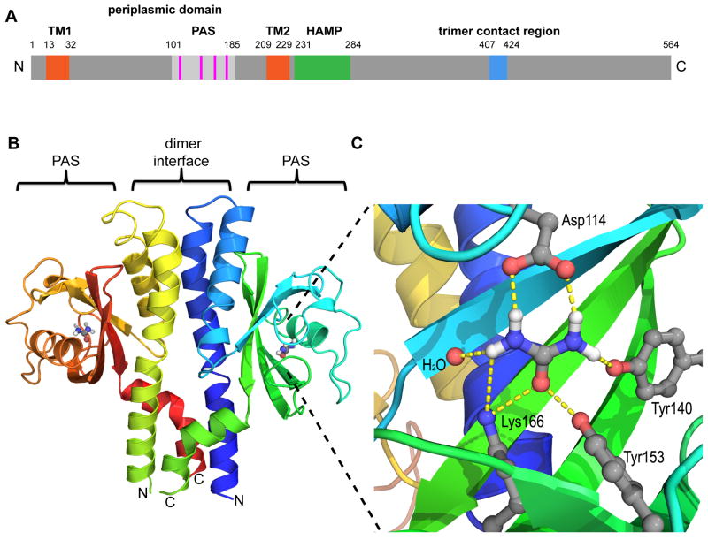
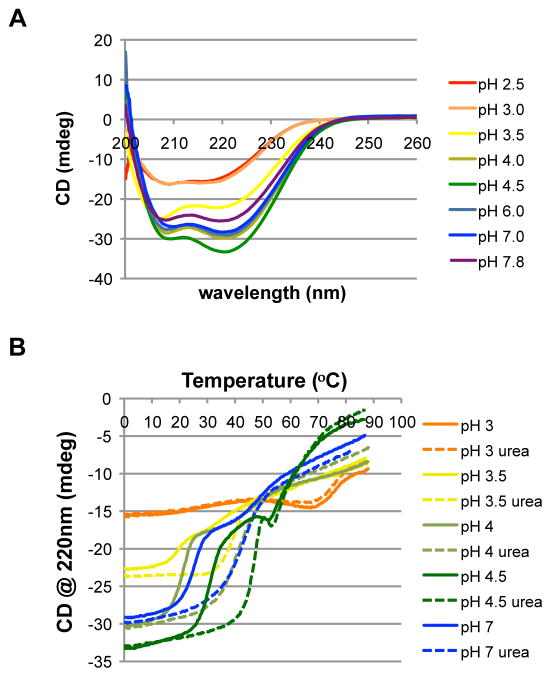
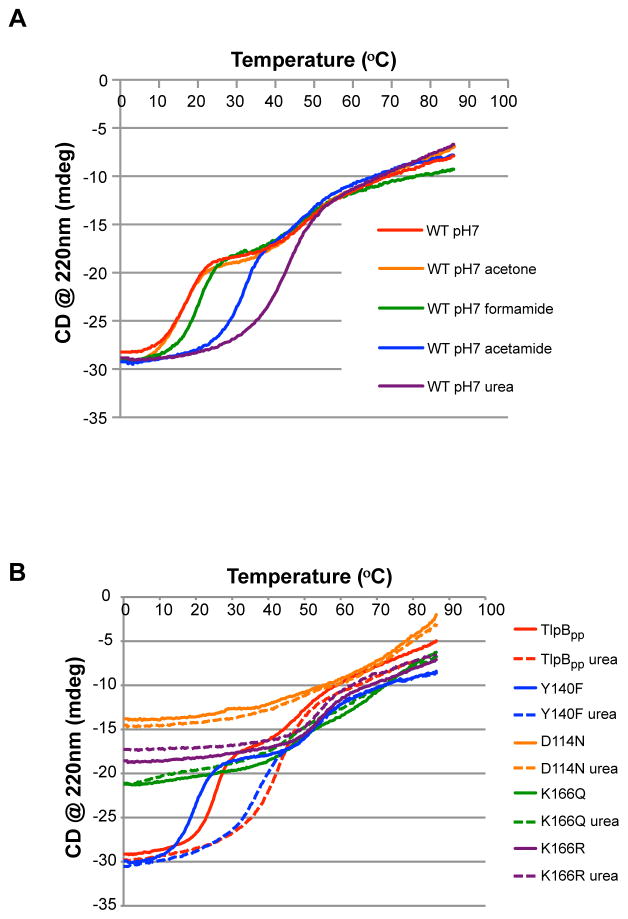
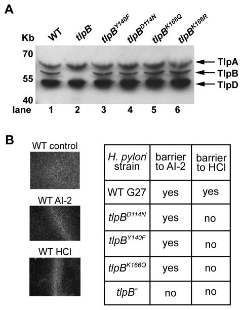
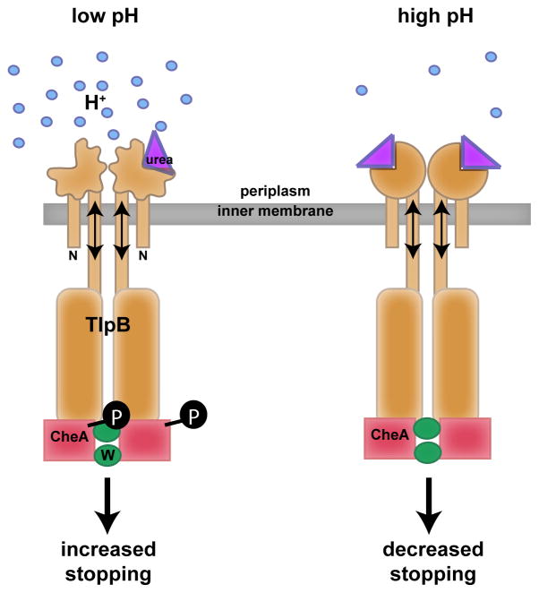
pH sensing is crucial for survival of most organisms, yet the molecular basis of such sensing is poorly understood. Here, we present an atomic resolution structure of the periplasmic portion of the acid-sensing chemoreceptor, TlpB, from the gastric pathogen Helicobacter pylori. The structure reveals a universal signaling fold, a PAS domain, with a molecule of urea bound with high affinity. Through biophysical, biochemical, and in vivo mutagenesis studies, we show that urea and the urea-binding site residues play critical roles in the ability of H. pylori to sense acid. Our signaling model predicts that protonation events at Asp114, affected by changes in pH, dictate the stability of TlpB through urea binding.


Figures

Figure 1
Figure 1
TlpBpp forms a dimer that contains urea-binding PAS domains. (A) Schematic of estimated SS1 TlpB domains. TM (orange), trans-membrane region; PAS (light gray), per-ARNT-Sim domain; HAMP (green), Histidine kinases, Adenylyl cyclases, Methyl binding proteins, Phosphatases domain; trimer contact region (blue), chemoreceptor trimer of dimers contact region. The periplasmic domain is from amino acid ~32–209. (B) Ribbon diagram of TlpBpp homodimer and gray/blue/red/white urea molecules, with chain color gradation ranging from N-terminus (blue) to C-terminus (green) for the monomer on the right and N-terminus (green) to C-terminus (red) for the monomer on the left. For orientation, the bacterial inner membrane would be below the lower part of the protein model diagram. (C) Diagram of the TlpBpp urea binding site including hydrogen bonds (dashed lines) between urea (gray, white, blue and red) and the surrounding residues and water molecule. Oxygen atoms are shown as red spheres, nitrogen as blue and hydrogen as white. See also Table 1 and Figure S1.
Figure 2
Figure 2
Urea thermally stabilizes TlpB in a pH dependent manner. (A) Circular dichroism spectra of TlpBpp with endogenous urea at multiple pHs reveals α-helical nature (spectra taken at 2ºC). TlpB concentration was 1 μM. (B) CD thermal melts of TlpBpp at multiple pHs. Dashed lines refer to samples with 5mM exogenously added urea. TlpB concentration for each curve was 1 μM. See also Figure S2.
Figure 3
Figure 3
Urea stabilizes TlpBpp specifically, and mutations in the urea binding pocket of TlpBpp abrogate the structure of TlpBpp and urea’s thermal stability effect. (A) CD thermal melts of TlpBpp with urea-like compounds (acetone, formamide and acetamide) added or urea added exogenously (all chemicals added at 5mM). TlpB concentration for each curve was 1 μM. (B) CD thermal melts of TlpBpp wild type (TlpBpp), Y140F, K166Q, K166R and D114N with urea (dashed lines) or without 5mM urea (solid lines). All samples were at pH 7. See also Figure S3.
Figure 4
Figure 4
H. pylori strains containing TlpB with urea binding point mutations show normal protein expression levels and remain responsive to AI-2. (A) Western blot of H. pylori G27 wild type (WT), tlpB knock out (tlpB) or the four point mutants (tlpBY140F, tlpBD114N, tlpBK166Q, tlpBK166R) using an antibody to the common, cytoplasmic region among all four chemoreceptors. (B) Results for the chemotaxis barrier assay. Images on the left show that wild type (WT) H. pylori respond to AI-2 and HCl by forming a barrier. The chart on the right shows responses of wild type, the tlpB strain and three of the TlpB urea binding mutant strains to AI-2 and HCl in the barrier assay. Images of AI-2 response to the five strains can be found in Figure S4A. See also Movie S1 and Figure S4.
Figure 5
Figure 5
TlpB mutants in the urea binding site have reduced or no response to acid while a reengineered urea-mimetic mutant remains responsive to acid. (A) Video chemotaxis assay tracks of individual wild type H. pylori shown for 10sec videos with no treatment (control) or HCl treatment. The bacteria showed more linear motion in the control compared to more curving and pausing in the presence of HCl. (B) Results of the chemotaxis video assay for a range of different HCl concentrations using wild type G27 H. pylori. Each dot represents a single video with the stops/sec of each track averaged. (C) Results of the chemotaxis video assay performed with wild type H. pylori (WT), cheA knock out (cheA−), tlpB knock out (tlpB−), tlpBD114N, tlpBY140F and tlpBK166Q with and without HCl treatment. The HCl treatment results are shown as red dots, the control treatments as black dots. (D) Results of the chemotaxis video assay performed with wild type H. pylori (WT) and tlpBK166R. An asterisk in C or D indicates that the pair is statistically significantly different using ANOVA with Tukey’s pairwise comparisons (Alpha = 0.01).
Figure 6
Figure 6
Model for how TlpB senses acid. In low pH conditions (shown on the left), TlpB’s periplasmic domain is in a “relaxed” or expanded state due to decreased hydrogen bonding to urea and consequent lowered urea binding affinity. However, in high or more neutral pH conditions, TlpB’s periplasmic domain is in a “tense” or condensed state with increased urea binding affinity. The state of the periplasmic domain is relayed through the transmembrane region which affects CheA’s phosphorylation state, ultimately affecting the flagellar motor and dictating stopping behavior. TlpB dimer (orange), CheA (red), CheW (green), urea (purple), protons (blue), phosphate (black).

Similar articles

Cited by

References

    1. Bowie JU, Pakula AA, Simon MI. The three-dimensional structure of the aspartate receptor from Escherichia coli. Acta Crystallogr D Biol Crystallogr. 1995;51:145–154. - PubMed
    1. Bowie JU, Sauer RT. Equilibrium dissociation and unfolding of the Arc repressor dimer. Biochemistry. 1989;28:7139–7143. - PubMed
    1. Briegel A, Ortega DR, Tocheva EI, Wuichet K, Li Z, Chen S, Muller A, Iancu CV, Murphy GE, Dobro MJ, et al. Universal architecture of bacterial chemoreceptor arrays. Proc Natl Acad Sci U S A. 2009;106:17181–17186. - PMC - PubMed
    1. Celej MS, Dassie SA, Freire E, Bianconi ML, Fidelio GD. Ligand-induced thermostability in proteins: thermodynamic analysis of ANS-albumin interaction. Biochim Biophys Acta. 2005a;1750:122–133. - PubMed
    1. Celej MS, Fidelio GD, Dassie SA. Protein Unfolding Coupled to Ligand Binding: Differential Scanning Calorimetry Simulation Approach. J Chem Ed. 2005b;82:87–92.

Publication types

MeSH terms

LinkOut - more resources