. 2012 Jul 13;423(4):763-9.
doi: 10.1016/j.bbrc.2012.06.033. Epub 2012 Jun 15.

Hydrogen peroxide induces stress granule formation independent of eIF2α phosphorylation

Affiliations

Hydrogen peroxide induces stress granule formation independent of eIF2α phosphorylation

Mohamed M Emara et al. Biochem Biophys Res Commun. .

Abstract

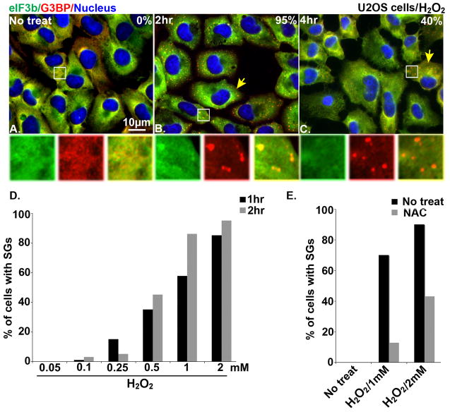
In cells exposed to environmental stress, inhibition of translation initiation conserves energy for the repair of cellular damage. Untranslated mRNAs that accumulate in these cells move to discrete cytoplasmic foci known as stress granules (SGs). The assembly of SGs helps cells to survive under adverse environmental conditions. We have analyzed the mechanism by which hydrogen peroxide (H(2)O(2))-induced oxidative stress inhibits translation initiation and induces SG assembly in mammalian cells. Our data indicate that H(2)O(2) inhibits translation and induces the assembly of SGs. The assembly of H(2)O(2)-induced SGs is independent of the phosphorylation of eIF2α, a major trigger of SG assembly, but requires remodeling of the cap-binding eIF4F complex. Moreover, H(2)O(2)-induced SGs are compositionally distinct from canonical SGs, and targeted knockdown of eIF4E, a protein required for canonical translation initiation, inhibits H(2)O(2)-induced SG assembly. Our data reveal new aspects of translational regulation induced by oxidative insults.


Figures

Fig. 1
Fig. 1
H2O2 induces SGs in U2OS cells. Immunofluorescence microscopy of U2OS cells that were left untreated (A) or treated with 1 mM H2O2 for 2 hr (B) or 4 hr (C) and then stained with SG markers [eIF3b (green) and G3BP (red)]. Nuclei are stained with Hoechst. Insets show enlarged views of individual and merged channels. Yellow arrows point out cells containing SGs. (D) Percentage of U2OS cells with G3BP-positive SGs after 2 hr treatment with 0.05 mM, 0.1 mM, 0.25 mM, 0.5 mM, 1 mM, and 2 mM H2O2. (E) The effect of NAC on H2O2-induced SG assembly. U2OS cells were treated with 50 mM of NAC for 1 hr then treated with 1 mM or 2 mM H2O2 for 2hr before processing for immunofluorescence microscopy.
Fig. 2
Fig. 2
H2O2 induces the assembly of non-canonical SGs. Immunofluorescence microscopy showing non-treated U2OS cells (A; No treat), cells treated with 250 μM sodium arsenite (B; SA) or 2 mM H2O2 (C; H2O2), or cells treated with H2O2 followed emetine treatment (D; H2O2+Em). Yellow arrows indicate SGs stained with SG markers [TIA-1 (green), PABP (red) and eIF4E (blue)]. Insets show enlarged views of individual and merged channels. (E) Percentage of cells with SA- or H2O2-induced SGs that contain the indicated protein markers. The average percentage of cells with SGs is shown (n=3). Error bars indicate the standard deviation. *=P values, that were calculated by comparing the percentage of cells with SGs in SA- and H2O2-treated cells (PABP, p=0.04; eIF4E, p=0.0006; eIF4G, p=0.002; and eIF3b, p=0.004). No other protein markers (G3BP, FXR1, TIA-1, and HuR) showed statistically significant differences. (F) Percentage of cells with SA- and H2O2-induced SGs before and after treatment with emetine. Error bars indicate the standard deviations of the mean (n=3). *=P value<0.005 when comparing the percentages of SGs in SA- or H2O2-treated cells with those treated with emetine.
Fig. 3
Fig. 3
H2O2 induces SGs independent of eIF2α phosphorylation. (A) Western blot analysis of phospho-eIF2α (upper panel) in U2OS cells treated with different concentrations of H2O2 (1, 2, 3, 4, and 5 mM) as well as 250 μM sodium arsenite (SA). TIA-1 (lower panels) was used as a loading control. (B–I) Immunofluorescence microscopy showing SG assembly induced by H2O2 in wild type (WT MEFs) and S51A eIF2α mutant MEFs (S51A MEFs). Yellow arrows indicate representative G3BP+ SGs (green) in untreated U2OS cells (B and F), or cells treated with 200 μM sodium arsenite (SA) (C and G), 50 nM pateamine A (PA) (D and H), and 2 mM H2O2 (E and I). Nuclei are stained with Hoechst.
Fig. 4
Fig. 4
H2O2 induces SGs by targeting eIF4F. (A) H2O2 causes eIF4F complex disruption and enhances eIF4E:4E-BP1 interactions. U2OS cells without (No treat) or with drug treatment (sodium aresenite (SA) and H2O2) were assembled on m7GTP-Sepharose as described in [18]. Loading control (Input) and the m7GTP-bound proteins (m7GTP-beads) were analyzed by Western Blotting using antibodies against eIF4G, eIF4A, eIF4E and 4E-BP1. (B) Western blot analysis showing the knockdown efficiency of eIF4E in U2OS cells transfected with control siRNA (si-Ctrl) or eIF4E-specific siRNA (si-4E). TIA-1 was used as a loading control. (C) Immunofluorescence microscopy showing SG assembly in U2OS cells after eIF4E depletion. Cell treated with the indicated siRNAs were left untreated (No treat, upper panels) or treated with 250 μM SA (middle panels), or 2 mM H2O2 (lower panels) and then stained with SG markers G3BP (red) and TIA-1 (green). Nuclei are stained with Hoechst. Insets show enlarged views of individual and merged channels. (D) Percentage of cells with SGs after eIF4E depletion. Error bars indicate the standard deviations of the mean (n=3, *p= 0.00036)

References

    1. Yamasaki S, Anderson P. Reprogramming mRNA translation during stress. Curr Opin Cell Biol. 2008;20:222–226. - PMC - PubMed
    1. Sonenberg N, Hinnebusch AG. Regulation of translation initiation in eukaryotes: mechanisms and biological targets. Cell. 2009;136:731–745. - PMC - PubMed
    1. Anderson P, Kedersha N. Stress granules: the Tao of RNA triage. Trends Biochem Sci. 2008;33:141–150. - PubMed
    1. Anderson P, Kedersha N. Stress granules. Curr Biol. 2009;19:397–408. - PubMed
    1. Anderson P, Kedersha N. RNA granules. J Cell Biol. 2006;172:803–808. - PMC - PubMed

Publication types