

A case of thoracolithiasis diagnosed thoracoscopically
- PMID: 22705577
- PMCID: PMC3397284
- DOI: 10.1016/j.ijscr.2012.05.006
A case of thoracolithiasis diagnosed thoracoscopically
Abstract
Introduction: Thoracolithiasis is quite rare with only 18 cases reported in the literature.
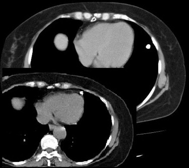
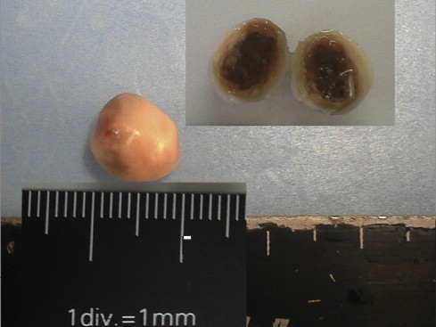
Presentation of case: The 65-year-old female was referred to us for the abnormality on the chest X-ray. The serial computed tomographic (CT) scans showed a mobile calcified nodule of about 10mm in diameter. Exploratory thoracoscopy was performed. A 15mm pearl-like pleural stone was removed with uneventful postoperative course.
Discussion: Thoracolithiasis is very rare, and its etiology as well as its epidemiology still remains to be unclear. Preoperative diagnosis is always difficult. However, specific radiological findings could let us recognize the probability of that lesion and avoid unnecessary major operation.
Conclusion: Thoracolithiasis should never be overlooked as a differential diagnosis in a pulmonary calcified nodule.
Copyright © 2012 Surgical Associates Ltd. Published by Elsevier Ltd. All rights reserved.
Figures
References
-
- Kinoshita F., Saida Y., Okajima Y., Honda S., Sato T., Hayashibe A. Thoracolithiasis: 11 cases with a calcified intrapleural loose body. J Thorac Imaging. 2010;25(1):64–67. - PubMed
-
- Dias A.R., Zerbini E.J., Curi N. Pleural stone. A case report. J Thorac Cardiovasc Surg. 1968;56(1):120–122. - PubMed
-
- Dadrich M., Schneider T., Puderbach M., Kauczor H.U., Heussel C.P. Moving pleural mass. Med Klin Intensivmed Notfmed. 2012 - PubMed
-
- Kosaka S., Kondo N., Sakaguchi H., Kitano T., Harada T., Nakayama K. Thoracolithiasis. Jpn J Thorac Cardiovasc Surg. 2000;48(5):318–321. - PubMed
-
- Bolca C., Trahan S., Frechette E. Intrapleural thoracolithiasis: a rare intrathoracic pearl-like lesion. Thorac Cardiovasc Surg. 2011;59(7):445–446. - PubMed
LinkOut - more resources
Full Text Sources
Research Materials
