. 2012 Nov;19(11):1847-55.
doi: 10.1038/cdd.2012.69. Epub 2012 Jun 15.

Differential regulation of DNA damage response activation between somatic and germline cells in Caenorhabditis elegans

Affiliations

Differential regulation of DNA damage response activation between somatic and germline cells in Caenorhabditis elegans

J Vermezovic et al. Cell Death Differ. 2012 Nov.

Abstract

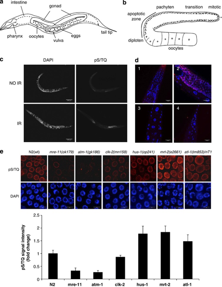
The germline of Caenorhabditis elegans is a well-established model for DNA damage response (DDR) studies. However, the molecular basis of the observed cell death resistance in the soma of these animals remains unknown. We established a set of techniques to study ionizing radiation-induced DNA damage generation and DDR activation in a whole intact worm. Our single-cell analyses reveal that, although germline and somatic cells show similar levels of inflicted DNA damage, somatic cells, differently from germline cells, do not activate the crucial apical DDR kinase ataxia-telengiectasia mutated (ATM). We also show that DDR signaling proteins are undetectable in all somatic cells and this is due to transcriptional repression. However, DNA repair genes are expressed and somatic cells retain the ability to efficiently repair DNA damage. Finally, we demonstrate that germline cells, when induced to transdifferentiate into somatic cells within the gonad, lose the ability to activate ATM. Overall, these observations provide a molecular mechanism for the known, but hitherto unexplained, resistance to DNA damage-induced cell death in C. elegans somatic cells. We propose that the observed lack of signaling and cell death but retention of DNA repair functions in the soma is a Caenorhabditis-specific evolutionary-selected strategy to cope with its lack of adult somatic stem cell pools and regenerative capacity.


Figures

Figure 1
Figure 1
Somatic post-mitotic cells of C. elegans do not show IR-induced pS/TQ signals upon IR. (a) Schematic representation of C. elegans anatomy, outlining the main structures in the body: pharynx, intestine and components of the reproductive system. (b) Schematic representation of the germline. Distinct phases of germ cells differentiation into oocytes are shown. The asterisk indicates the distal end of the germline. Cell death occurs only in the outlined (‘apoptotic zone') pachytene subphase of meiosis. (c) pS/TQ staining of intact wild-type (N2) worms treated with 180 Gy of IR and stained 1 h post treatment. Scale bar, 100 μm. (d) Close up and merged (blue: DAPI; red: pS/TQ) images of different regions of worm body (1: head, 2: mid-body, 3: large nuclei of the hypodermis, 4: tail), showing the absence of pS/TQ signal in somatic cells. Scale bar, 20 μm. (e) Representative images of pS/TQ staining of meiotic nuclei of the indicated genotypes after IR treatment. Histogram represents fold change in the mean intensity of the pS/TQ signal for the indicated genotype, normalized to the signal intensity of irradiated wild-type (N2) worms. At least 10 gonads per strain were analyzed. Error bars represent the mean±S.E.M. Scale bar, 5 μm
Figure 2
Figure 2
Both somatic and germline cells show the presence of DNA damage upon IR, but only germline cells show DDR activity. (a) Mid-body of wild-type (N2) worms showing both germline and somatic cells, stained by TUNEL assay for the presence of DNA breaks. Scale bar, 20 μm. (b) qRT-PCR performed on wild type (N2) and (glp-1) IR-treated worms, with primers specific for egl-1 gene, showing its induction only in the irradiated wild-type worms. mRNA levels were normalized to the value of wild type not irradiated worms. Error bars represent the mean±S.D. for a representative experiment performed in triplicate. (c) pS/TQ staining of cku-70(tm1724) and lig-4(ok716) IR-treated mutants show DDR signaling only in the germline. The asterisk indicates distal end of the germline. (d) DIC images and pS/TQ staining of four distinct developmental larval stages (L1–L4) of irradiated wild-type (N2) worms. Scale bar, 200 μm
Figure 3
Figure 3
DDR genes are not expressed in the worm soma. (a) Differential interference contrast and fluorescent microscopy of wild type and transgenic animals expressing: HUS-1::GFP(opIs334), RPA-1::YFP(opIs263), MRE-11::YFP (opIs239) and CEP-1::GFP(gtIs1), RPA-1 is expressed in all worm cells, while MRE-11, HUS-1 and CEP-1 are expressed in germline cells only (close ups of the somatic cells and germline are indicated by a white arrow). The asterisk indicates distal end of the germline. Scale bar for whole-worm images 100 μm, and 50 μm for close ups. (b) qRT-PCR performed on wild type (N2) and (glp-1) worms with primers specific for the indicated DDR genes. Error bars represent the mean±S.D. for a representative experiment performed in triplicate
Figure 4
Figure 4
Worm somatic cells show DNA repair proficiency. (a) Fully somatic (glp-1) worms were irradiated with the indicated dose of IR and single-worm PCR was performed in quadruplicate, per irradiation point, either immediately after irradiation (T0) or 24 h after irradiation (T24). Bars on the histogram are mean values (for quadruplicate) of the 11 kb PCR product quantity normalized to the 1 kb PCR product quantity, and to the values of not irradiated wild-type worms. Error bars are mean±S.E.M. (b) glp-4(bn2) and glp-4(bn2) lig-4(ok716) mutants were irradiated with 100 Gy of X-rays and probed for DNA damage by PCR assay immediately (T0) or 24 h after irradiation (T24). Histogram represents quantification of PCR products for the biological triplicate
Figure 5
Figure 5
Somatic cells misexpressing germline genes do not activate DDR, while germline cells transdifferentiated into somatic cells lose DDR signaling. (a) lin-35(n745) and lin-15b(n744) mutants were exposed to IR and co-stained with pS/TQ and germline specific marker PGL-1. (b) Wild-type (N2) worms were treated with 10 mM VPA from L1 to the adult stage, exposed to IR and stained for pS/TQ and acetylated histone H4. Somatic cells that show the absence of pS/TQ signal are outlined with white circles. (c) mex-3(or20) gld-1(q485) unc-119::gfp mutant stained for pS/TQ (red), myosin (magenta) and neurons express unc-119::GFP (green). White arrows point to the nuclei of neuronal cells expressing unc-119::GFP, yellow arrows point to the nuclei of muscle cells stained with myosin. Scale bars on all images, 10 μm

References

    1. Matsuoka S, Ballif BA, Smogorzewska A, McDonald ER, Hurov KE, Luo J, et al. ATM and ATR substrate analysis reveals extensive protein networks responsive to DNA damage. Science. 2007;316:1160–1166. - PubMed
    1. Branzei D, Foiani M. Regulation of DNA repair throughout the cell cycle. Nat Rev Mol Cell Biol. 2008;9:297–308. - PubMed
    1. Gartner A, Milstein S, Ahmed S, Hodgkin J, Hengartner MO. A conserved checkpoint pathway mediates DNA damage--induced apoptosis and cell cycle arrest in C. elegans. Mol Cell. 2000;5:435–443. - PubMed
    1. Kim ST, Lim DS, Canman CE, Kastan MB. Substrate specificities and identification of putative substrates of ATM kinase family members. J Biol Chem. 1999;274:37538–37543. - PubMed
    1. Carballo JA, Johnson AL, Sedgwick SG, Cha RS. Phosphorylation of the axial element protein Hop1 by Mec1/Tel1 ensures meiotic interhomolog recombination. Cell. 2008;132:758–770. - PubMed

Publication types

MeSH terms

Substances