. 2012 Jun 15;31(14):3169-82.
doi: 10.1038/emboj.2012.165.

Glutathione redox potential in the mitochondrial intermembrane space is linked to the cytosol and impacts the Mia40 redox state

Affiliations

Glutathione redox potential in the mitochondrial intermembrane space is linked to the cytosol and impacts the Mia40 redox state

Kerstin Kojer et al. EMBO J. .

Abstract

Glutathione is an important mediator and regulator of cellular redox processes. Detailed knowledge of local glutathione redox potential (E(GSH)) dynamics is critical to understand the network of redox processes and their influence on cellular function. Using dynamic oxidant recovery assays together with E(GSH)-specific fluorescent reporters, we investigate the glutathione pools of the cytosol, mitochondrial matrix and intermembrane space (IMS). We demonstrate that the glutathione pools of IMS and cytosol are dynamically interconnected via porins. In contrast, no appreciable communication was observed between the glutathione pools of the IMS and matrix. By modulating redox pathways in the cytosol and IMS, we find that the cytosolic glutathione reductase system is the major determinant of E(GSH) in the IMS, thus explaining a steady-state E(GSH) in the IMS which is similar to the cytosol. Moreover, we show that the local E(GSH) contributes to the partially reduced redox state of the IMS oxidoreductase Mia40 in vivo. Taken together, we provide a comprehensive mechanistic picture of the IMS redox milieu and define the redox influences on Mia40 in living cells.


Conflict of interest statement

The authors declare that they have no conflict of interest.

Figures

Figure 1
Figure 1
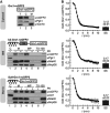
Establishment of an assay to dynamically monitor the glutathione redox potential. (A) Properties of the Grx1-roGFP2 sensor. WT yeast cells expressing Grx1-roGFP2 were grown to mid-log phase in galactose-containing media. Excitation spectra (λemission=511 nm) were recorded on untreated cells (steady state=SS), and on cells pretreated with DTT or diamide (Dia). In parallel, the sensor redox states were analysed by alkylation shift assays (inset). (B) Ratiometric analysis of the steady state of the Grx1-roGFP2 sensor in the cytosol of S. cerevisiae. Excitation spectra were obtained as described in (A). The ratio of the intensities at 405 and 488 nm was formed for the steady state, the DTT and the diamide value. (C) Steady state of the Grx1-roGFP2 sensor in the cytosol of S. cerevisiae. The steady-state ratio from (B) was transformed to the degree of sensor oxidation—OxD (for calculation see Materials and methods) with an OxD=1 for the fully oxidized probe; OxD=0 for the fully reduced probe. (D) Scheme for the set-up of an oxidant recovery experiment. Yeast cells were incubated with 20 mM diamide for 5 min at 30°C under constant agitation, the oxidant was removed by washing twice with measurement buffer, and the recovery of the respective Grx1-roGFP2 sensors was monitored using a fluorescence spectrometer. Excitation spectra were obtained at different times after diamide washout using an emission wavelength of 511 nm. (E) Excitations spectra of an oxidant recovery experiment using Grx1-roGFP2. WT yeast cells expressing Grx1-roGFP2 were grown to mid-log phase in SC galactose media, and treated according to the protocol described in (D). (F) Recovery assay after oxidative shock. As (E) except that the intensities at the excitation wavelengths 405 and 488 nm were normalized to OxD values. The experiment could be repeated with the same sample (recovery 1, recovery 2). In parallel to recovery 1, the sensor redox states were analysed by alkylation shift assays (inset). (G) False colour fluorescence microscopy images of the recovery of the cytosolic redox sensor after oxidative shock in yeast cells.
Figure 2
Figure 2
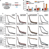
EGSH[cytosol], EGSH[IMS] and EGSH[matrix] recover fast and with similar kinetics to their steady-state values after oxidative shock. (A) Mitochondria-targeted constructs of Grx1-roGFP2 and their subcellular localization. The white box represents the Grx1-roGFP2 protein, the helix represents the amphipathic helix required for matrix targeting, and the grey box represents the hydrophobic sorting domain required for IMS targeting. The targeting sequences of the mitochondrial proteins subunit 9 of the ATPase (Su9, aa 1–69) and cytochrome b2 (aa 1–86) were fused to Grx1-roGFP2. The subcellular localization of Grx1-roGFP2, Su9-Grx1-roGFP2 and b2-Grx1-roGFP2 was assessed in WT yeast cells (BY4742) expressing Grx1-roGFP2, Su9-Grx1-roGFP2 and b2-Grx1-roGFP2. Cells were grown to mid-log phase in SLac+0.1% galactose media, they were lysed and fractionated, and fractions were analysed by SDS–PAGE and immunoblotting using antibodies directed against roGFP2, 3-phosphoglycerate kinase (Pgk1) and actin (Act1; both cytosol), the mitochondrial ribosomal protein Mrpl36 (matrix), Mia40 (IMS), or the subunit of the translocase of the outer membrane Tom70 (OMM). Cells were fractionated into post-mitochondrial supernatant (C) and mitochondria (M). Mitoplasts (MP) were generated from mitochondria by hypo-osmotic swelling. Mitochondria were also lysed with Triton X-100 (TX-100), and all fractions were treated with proteinase K (PK) as indicated. (B) Recovery of the redox states of Grx1-roGFP2 probes in different compartments after diamide washout. WT yeast cells expressing Grx1-roGFP2, Su9-Grx1-roGFP2 and b2-Grx1-roGFP2 were analysed as described in Figure 1F (growth on galactose as carbon source). In addition, the redox states of the sensors at steady state were obtained as described in Figure 1C. Reported values are the mean of three independent experiments. Error bars are the means±s.d.
Figure 3
Figure 3
The cytosolic glutathione reductase system is the major determinant of EGSH[IMS]. (A) Scheme of the enzymes analysed in these experiments. (B) Steady states of the cytosolic and mitochondrial sensors (Grx1-roGFP2, Su9-Grx1-roGFP2, b2-Grx1-roGFP2) in yeast mutants of the glutathione system. Experiment was performed as described in Figure 1C. (CF) Recovery kinetics after diamide shock on mutants of the glutathione reductase system. WT yeast cells were compared to cells from Δglr1 (C), Δglr1+GLR1 (D) Δglr1+cyto-GLR1 (E) or Δzwf1 (F) strains, respectively. Cells expressing Grx1-roGFP2, Su9-Grx1-roGFP2 and b2-Grx1-roGFP2 were analysed as described in Figure 1F (growth on galactose as carbon source). Reported values are the mean of three independent experiments. Error bars are the means±s.d.
Figure 4
Figure 4
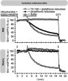
The IMS is accessible to exogenous GSH. Mitochondria from WT yeast cells harbouring either b2-Grx1-roGFP2 or Su9-Grx1-roGFP2 were isolated and analysed in a fluorescent spectrometer upon incubation with glutathione reductase, NADPH, GSH and Triton X-100 (TX-100) as indicated. After isolation of mitochondria, the respective probes are fully oxidized (=100% oxidized Grx1-roGFP2). The incubation with DTT at the end of the kinetics served as control for fully reduced proteins (=0% Grx1-roGFP2).
Figure 5
Figure 5
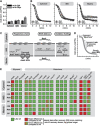
IMS redox pathways exert only a limited influence on EGSH[IMS]. (A) Steady states of the cytosolic and mitochondrial sensors (Grx1-roGFP2, Su9-Grx1-roGFP2, b2-Grx1-roGFP2) in wt cells grown on galactose (Gal) or glycerol (Glyc). Experiment was performed as described in Figure 1C. Reported values are the mean of three independent experiments. Error bars are the means±s.d. (B) Recovery kinetics after diamide shock in wt cells grown on galactose or glycerol. Cells expressing Grx1-roGFP2, Su9-Grx1-roGFP2 and b2-Grx1-roGFP2 were analysed as described in Figure 1F. Reported values are the mean of three independent experiments. Error bars are the means±s.d. (C) Scheme of the investigated IMS redox pathways. (D) Scheme depicting the three parameters to evaluate the recovery kinetics after oxidative shock: (i) apparent lag phase, (ii) recovery rate and (iii) steady-state OxD. For full description, see Supplementary Figure S2. (E) Qualitative representation of the results obtained from the analysis of (i) chemical treatments to inhibit complexes of the respiratory chain (antimycin A (AntA), potassium cyanide (KCN) to inhibit complexes III and IV, respectively, and paraquat to induce mitochondrial oxidative stress), (ii) mutants that result in defective respiratory chain complexes (Δrip1, Δcox17, Δnde1 and Δnde2), (iii) deletions of enzymes counteracting ROS (Δsod1, Δsod2, Δccp1) and (iv) strains that express Erv1 and Mia40 under the control of regulatable promoters. The full set of data is presented in Supplementary Figures S4–S8, respectively.
Figure 6
Figure 6
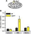
Porins control EGSH[IMS]. (A) Scheme of the strain used. (B) Steady states of the cytosolic and mitochondrial sensors (Grx1-roGFP2, Su9-Grx1-roGFP2, b2-Grx1-roGFP2) in Δpor1 cells. Experiment was performed as described in Figure 1C. Reported values are the mean of three independent experiments. Error bars are the means±s.d.
Figure 7
Figure 7
The in-vivo redox state of Mia40 is partially reduced. (A) The pathway for disulphide bond formation in the IMS. Reduced substrates are oxidized by Mia40 which in turn is re-oxidized by Erv1. From Erv1 electrons can be passed on directly to oxygen or to respiratory chain complex IV. (B) In-vivo redox state of Mia40 in wild-type yeast cells. Cells were precipitated with TCA. The resulting pellet was resuspended in a buffer containing 50 mM NEM. The samples were analysed on non-reducing SDS–PAGE followed by immunoblotting against Mia40. SS; steady state. Reported values are the mean of three independent experiments. Error bars are the means±s.d. (C) Erv1 levels in cells that express Erv1 under the control of a regulatable promoter. Cells were grown either in galactose or in glucose to raise or lower cellular Erv1 levels, respectively. Erv1 levels were tested by western blot analysis. (D) Redox state of Mia40 in cells with different Erv1 levels. As (B), except that cells expressed Erv1 under the control of a regulatable promoter. Cells were grown either in galactose or in glucose to raise or lower cellular Erv1 levels, respectively. Reported values are the mean of three independent experiments. Error bars are the means±s.d. (E) Redox state of Mia40 in Δrip1 and Δcox17 cells in the presence of 20 or 1% oxygen. As (B), except that the experiment was performed with Δrip1 and Δcox17 cells in the presence of different oxygen concentrations. The experiments have been performed three times independently. Error bars are the means±s.d. Figure source data can be found with the Supplementary data.
Figure 8
Figure 8
EGSH[IMS] is a determinant of the Mia40 redox state. (A) Scheme depicting the hypothetical influence of the glutathione pool on Mia40 thus giving rise to a partially reduced Mia40 at steady state. (B) Protein levels in wild type, Δglr1, GalL-Erv1 and GalL-Erv1/Δglr1 cells. (C) Redox states of Mia40 in wild type, Δglr1, GalL-Erv1 and GalL-Erv1/Δglr1 cells. Cells were analysed as described in Figure 7B. Reported values are the mean of three independent experiments. Error bars are the means±s.d. (D) Scheme for the set-up of an oxidant recovery experiment. Yeast cells were incubated with 20 mM diamide for 5 min at 30°C under constant agitation, the oxidant was removed by washing twice with measurement buffer, and the recovery of the Mia40 redox state was assessed at different times using the NEM alkylation-based gel shift assay. (E) Oxidant recovery kinetics of Mia40 in GalL-Erv1 and GalL-Erv1/Δglr1 cells. Cells were analysed as described in (C). Reported values are the mean of two independent experiments. Figure source data can be found with the Supplementary data.
Figure 9
Figure 9
Model for the dynamics, interplay and physiological impact of the cytosolic and mitochondrial glutathione pools. (A) Fast cross-talk via porins in the OMM takes place between the glutathione pools of the IMS and the cytosol. Because of this dynamic glutathione exchange the cytosolic glutathione reductase system exerts the major influence on the composition of the IMS glutathione pool. Although the matrix relies on glutathione delivery from the cytosol for replenishing its glutathione pool, EGSH[matrix] is maintained by matrix-localized reducing systems as GSSG cannot be exported from the matrix. (B) The oxidoreductase of the IMS Mia40 is in vivo partially reduced. Its redox state is maintained by reducing influences from the local glutathione pool (and newly imported reduced substrates), and oxidizing influences from the sulphhydryloxidase Erv1. In-vivo Erv1 can shuttle its electrons either directly to oxygen or via the respiratory chain. This latter pathway might only be required under conditions of low oxygen.

Similar articles

Cited by

References

    1. Antos N, Budzinska M, Kmita H (2001) An interplay between the TOM complex and porin isoforms in the yeast Saccharomyces cerevisiae mitochondria. FEBS Lett 500: 12–16 - PubMed
    1. Appenzeller-Herzog C (2011) Glutathione- and non-glutathione-based oxidant control in the endoplasmic reticulum. J Cell Sci 124: 847–855 - PubMed
    1. Ballatori N, Krance SM, Marchan R, Hammond CL (2009) Plasma membrane glutathione transporters and their roles in cell physiology and pathophysiology. Mol Aspects Med 30: 13–28 - PMC - PubMed
    1. Banhegyi G, Lusini L, Puskas F, Rossi R, Fulceri R, Braun L, Mile V, di Simplicio P, Mandl J, Benedetti A (1999) Preferential transport of glutathione versus glutathione disulfide in rat liver microsomal vesicles. J Biol Chem 274: 12213–12216 - PubMed
    1. Bien M, Longen S, Wagener N, Chwalla I, Herrmann JM, Riemer J (2010) Mitochondrial disulfide bond formation is driven by intersubunit electron transfer in Erv1 and proofread by glutathione. Mol Cell 37: 516–528 - PubMed

MeSH terms

Substances