. 2012 Sep-Dec;129(9-12):308-23.
doi: 10.1016/j.mod.2012.06.002. Epub 2012 Jun 15.

Delayed fusion and altered gene expression contribute to semicircular canal defects in Chd7 deficient mice

Affiliations

Delayed fusion and altered gene expression contribute to semicircular canal defects in Chd7 deficient mice

Elizabeth A Hurd et al. Mech Dev. 2012 Sep-Dec.

Erratum in

Abstract

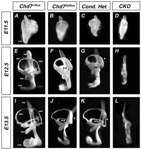
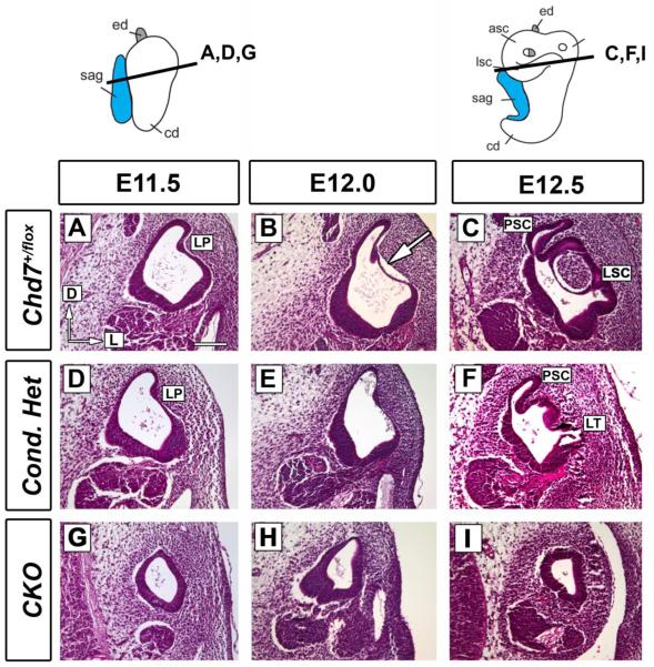
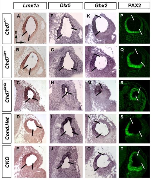
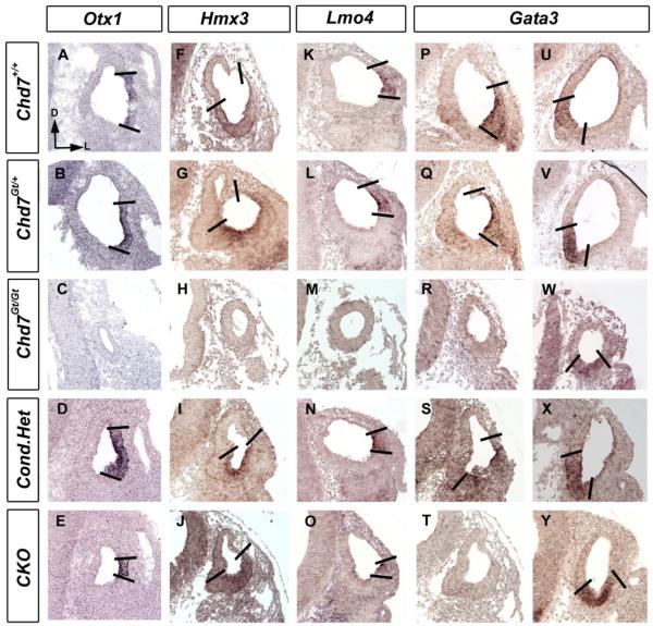
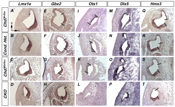
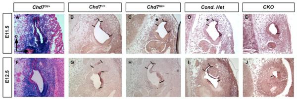
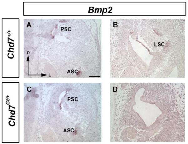
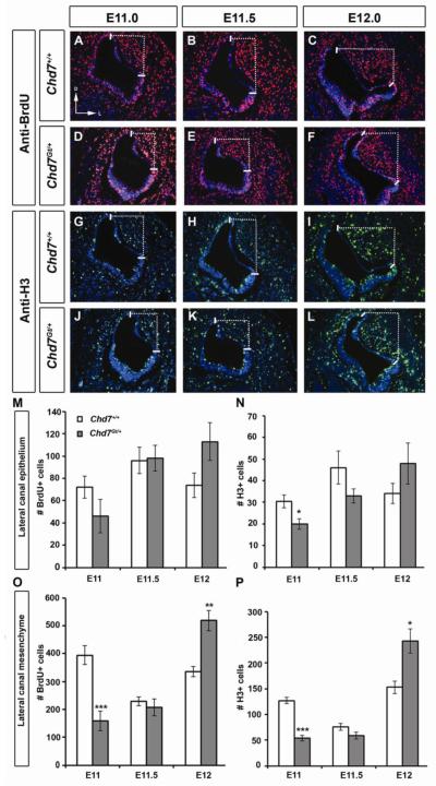
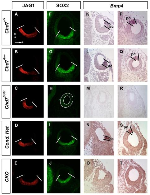
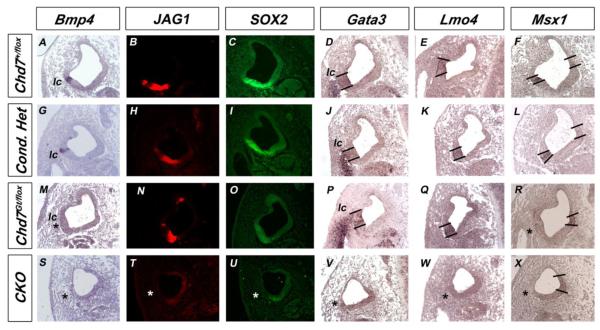
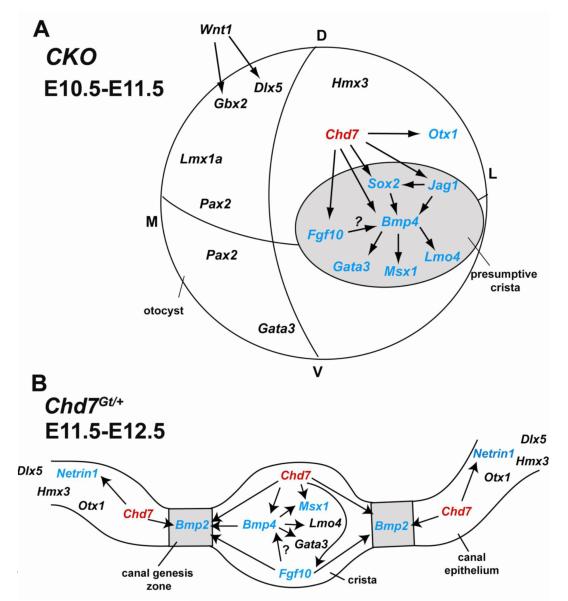
Proper morphogenesis of inner ear semicircular canals requires precise regulation of cellular proliferation, epithelial-to-mesenchymal transition, and fusion of epithelial plates. Epigenetic regulation of these processes is not well understood, but is likely to involve chromatin remodeling enzymes. CHD7 is a chromodomain-containing, ATP dependent helicase protein that is highly expressed in the developing ear and is required for semicircular canal development in both humans and mice. Here we report that mice with heterozygous loss of Chd7 function exhibit delayed semicircular canal genesis, delayed Netrin1 expression and disrupted expression of genes that are critical for semicircular canal formation (Bmp2, Bmp4, Msx1 and Fgf10). Complete loss of Chd7 results in aplasia of the semicircular canals and sensory vestibular organs, with reduced or absent expression of Otx1, Hmx3, Jagged1, Lmo4, Msx1 and Sox2. Our results suggest that Chd7 may have critical selector gene functions during inner ear morphogenesis. Detailed analysis of the epigenetic modifications underlying these gene expression changes should provide insights into semicircular canal development and help in the design of therapies for individuals with inner ear malformations.


Figures

Figure 1
Figure 1. Posterior and lateral canal development is delayed in Chd7 mutant embryos
Chd7+/flox (A,E,I), Chd7Gt/flox (B,F,J), Chd7 conditional heterozygous (C,G,K) and Chd7 conditional null (D,H,L) inner ears after paint filling at E11.5-E13.5. (A-D) At E11.5, anterior and posterior pouches are observed in control (A), heterozygous (B) and conditional heterozygous (C) inner ears, but are hypoplastic in CKO (D). (E-G) At E12.5, formation of the posterior (*) and lateral (**) canals are delayed in heterozygous (F) and conditional heterozygous (G) ears. By E13.5, defects in posterior (*) and lateral (**) canals are observed in heterozygous (J) and conditional heterozygous (K) ears. No canals are present in CKO inner ears at either E12.5 (H) or E13.5 (L). Abbreviations: CKO= Chd7 conditional knockout (Foxg1-Cre;Chd7Gt/flox); asc, anterior semicircular canal; coc, cochlea; ed, endolymphatic duct; lsc, lateral semicircular canal; psc, posterior semicircular canal; sac, saccule.
Figure 2
Figure 2. Lateral semicircular canal fusion is delayed in Chd7 mutant embryos
Thin paraffin sections (7 μm) cut in the transverse plane at E11.5 (A, D, G), E12.0 (B, E, H) and E12.5 (C, F, I) were stained with Hematoxylin and Eosin at the level of the lateral canal. (A-C) Control (Chd7+/flox) semicircular canal epithelium detaches from the basement membrane (arrow) creating the lateral semicircular canal (LC). (D-F) Conditional heterozygous (Foxg1-Cre;Chd7+/flox ) inner ears exhibit delayed epithelial detachment (E vs. B), resulting in a truncated lateral semicircular canal (LT in F). (G-I) The CKO (Foxg1-Cre;Chd7Gt/flox ) vestibule displays a very small canal outpocketing (G) and no visible fusion plate or lateral semicircular canal (H-I). Orientation: dorsal is towards the top and lateral is to the right, as shown in (A). Other abbreviations: CKO, Chd7 conditional knockout (Foxg1-Cre;Chd7Gt/flox); LP, lateral pouch; LSC, lateral semicircular canal; LT, lateral truncation; PSC, posterior semicircular canal. Bar in A= 100 μm and applies to all panels.
Figure 3
Figure 3. Dorsomedial markers are unchanged in Chd7 mutant mice
In situ hybridization and immunofluorescence with probes against Lmx1a (A-E), Dlx5 (F-J), Gbx2 (K-O) and anti-PAX2 (P-T) shows unaltered expression patterns between E10.5 Chd7+/+ (A, F, K ,P), Chd7Gt/+ (B, G, L, Q), Chd7Gt/Gt (C, H, M, R), Foxg1-Cre;Chd7+/flox (D, I, N, S) and Foxg1-Cre;Chd7Gt/flox (E, J, O, T) otocysts. All sections are in the transverse orientation with dorsal towards the top as shown by the arrows in (A). Bold lines delimit expression domains. Abbreviation: CKO= Chd7 conditional knockout (Foxg1-Cre;Chd7Gt/flox).
Figure 4
Figure 4. Expression of genes in the lateral otocyst is reduced in Chd7 null mutant embryos
In situ hybridization with probes against Otx1 (A-E), Hmx3 (F-J), Lmo4 (K-O) and Gata3 (P-Y) shows unchanged expression in E10.5 Chd7+/+ (A, F, K, P, U), Chd7Gt/+ (B, G, L, Q, V) and Foxg1-Cre;Chd7+/flox (D, I, N, S, X). Otx1 (E) and dorsal Gata3 (T) expression are reduced in CKO embryos, whereas Hmx3 (J), Lmo4 (O), and ventral Gata3 (Y) expression are preserved. Hypoplastic Chd7Gt/Gt otocysts show no Otx1, Hmx3, Lmo4 or dorsal Gata3 expression, but maintain ventral Gata3 expression (W). All sections are in the transverse orientation with dorsal towards the top as shown by the arrows in (A). Bold lines delimit expression domains. Abbreviation: CKO = Chd7 conditional knockout (Foxg1-Cre;Chd7Gt/flox).
Figure 5
Figure 5. Otx1 expression is reduced in Chd7 mutant embryos
In situ hybridization with probes against Lmx1a (A-D), Gbx2 (E-H), Otx1 (I-L), Dlx5 (M-P) and Hmx3 (Q-T) show unaltered expression patterns in E11.5 Chd7 +/flox (A, E, I, M, Q), conditional heterozygous (B, F, J, N, R) and Chd7Gt/+ (C, G, K, O, S) ears. Lmx1a (D), Gbx2 (H), Hmx3 (T) and medial Dlx5 (P) expression were preserved in Chd7 CKO (Foxg1-Cre;Chd7Gt/flox) ears. Lateral Dlx5 (P) and Otx1 (L) expression were absent from CKO inner ears. All sections are in the transverse orientation at the level of the lateral semicircular canal. Bold lines delimit expression domains. Abbreviation: CKO= Chd7 conditional knockout (Foxg1-Cre;Chd7Gt/flox).
Figure 6
Figure 6. Medial Netrin1 expression is delayed in Chd7 mutant semicircular canal primordia
Transverse sections at the level of the lateral semicircular canal primordia at E11.5 (A-E) and E12.5 (F-J) were stained for β-galactosidase activity (A, F) or Netrin1 mRNA (B-E,G-J). (A, F) β-galactosidase activity shows Chd7 promoter activity within the periotic mesenchyme surrounding the semicircular canals and within the primordial canal epithelium. In situ hybridization shows medial (*) and lateral expression of Netrin1. Medial Netrin1 expression (*) is delayed in E11.5 germline and conditional Chd7 heterozygotes (C,D) . The entire Netrin1 expression domain is reduced in hypoplastic Foxg1-Cre;Chd7Gt/flox (I, J) inner ear. Bold lines delimit Netrin1 expression domains. Orientation for all panels is shown by the arrows in (A). Bar in A= 100 μm and applies to all panels. Abbreviation: CKO= Chd7 conditional knockout (Foxg1-Cre;Chd7Gt/flox).
Figure 7
Figure 7. Bmp2 expression is reduced in Chd7Gt/+ inner ears
In situ hybridization of E12.5 transverse sections of wild type (A,B) and Chd7Gt/+ (C, D) embryos shows selective absence of Bmp2 mRNA from the Chd7Gt/+ lateral canal region (D), despite normal expression in anterior and posterior regions (C). Orientation: dorsal towards the top and lateral to the right, as shown by the arrows in (A). Abbreviations: AC, Anterior semicircular canal; LC, Lateral semicircular canal; PC, posterior semicircular canal.
Figure 8
Figure 8. Proliferation is altered in the lateral canal epithelium and mesenchyme of Chd7Gt/+ embryos
Double immunofluorescence for anti-BrdU and anti-H3 of wild type (A-C, G-I) and Chd7Gt/+ (D-F, J-L) lateral semicircular canals at E11.0 (A,D,G,J), E11.5 (B,E,H,K) and E12.0 (C,F,I,L). Numbers of proliferating cells were counted in the lateral epithelium (between solid lines; M,N) and in the lateral periotic mesenchyme (within dotted lines; O,P). At E11.0, significant decrease in H3-positive cells in the Chd7Gt/+ epithelium (N) and mesenchyme (P), and BrdU-positive cells in Chd7Gt/+ mesenchyme (O) were observed. Increased BrdU-positive and H3-positive cells were observed in Chd7Gt/+ mesenchyme at E12.0 (N,P) compared to wild type controls. Sections are in the transverse plane, with dorsal towards the top as in (A). *P<0.05, **P<0.01, ***P<0.001.
Figure 9
Figure 9. Bmp4 and SOX2 in the developing crista are sensitive to Chd7 dosage
Immunofluorescence (A-J) or in situ hybridization (K-T) of E10.5 transverse sections. JAGGED1 is unaltered in Chd7 heterozygous (B, D) and Chd7 null (C, E) otocysts compared to control otocysts (A). SOX2 is unchanged in Chd7 heterozygous (G, I) and conditional null (J) otocysts but is absent from Chd7Gt/Gt (H) otocysts. Bmp4 is expressed within the anterior crista primordia (ac) in Chd7 heterozygous mutants (L, N) and is variably expressed in the posterior crista primordia (pc); some otocysts display positive Bmp4 expression in the posterior streak (Q, S) whereas it is absent from other otocysts. Bmp4 expression is absent from Chd7 null mutant otocyst (M, O). Bold lines delineate expression domains. Plane of section is shown by arrows in (A), with dorsal toward the top and lateral toward the right. Abbreviation: CKO= Chd7 conditional knockout (Foxg1-Cre;Chd7Gt/flox).
Figure 10
Figure 10. Expression of genes expressed in the lateral crista is altered in Chd7 mutant embryos
In situ hybridization (A, D-G, J-M, P-S, V-X) or immunofluorescence (B, C, H, I, N, O, T, U) of E11.5 transverse sections at the level of the lateral canal. Bmp4, JAG1, SOX2, Gata3, Lmo4 and Msx1 are present within the lateral crista primordium (lc) in conditional heterozygous mutants (G-L), but absent from CKO otocysts (* in S-X). Bmp4 expression is variably absent in Chd7Gt/flox otocysts (* in M), but JAG1 and SOX2 are preserved in adjacent sections (N, O). Expression of Gata3 and Lmo4 are unchanged in Chd7Gt/flox otocysts (P,Q), but lateral expression of Msx1 is absent (* in R). Bold lines delineate expression domains. Abbreviation: CKO= Chd7 conditional knockout (Foxg1-Cre;Chd7Gt/flox).
Figure 11
Figure 11. Bmp2, Bmp4, Otx1, Sox2 and Fgf10 in the otocyst are regulated by Chd7 in a dosage dependent manner
qRT-PCR analysis of total RNA from E10.5 Chd7+/+, Chd7Gt/+ and Chd7Gt/Gt microdissected inner ears shows dosage sensitive reductions with loss of Chd7. Threshold cycles from the experimental genes were normalized to Gapdh, and the Chd7Gt/+ and Chd7Gt/Gt were compared to wild type (Chd7+/+). Error bars are standard error of the mean, n=3 for each genotype. *P<0.05
Figure 12
Figure 12. Model of Chd7 function in genetic cascades that regulate lateral semicircular canal development
(A) Conditional loss of Chd7 using Foxg1-Cre results in reduced expression of Bmp4, Sox2, Otx1, Jag1, Fgf10, Gata3, Msx1 and Lmo4 in the dorsal otocyst and presumptive crista (grey circle). (B) Heterozygous loss of Chd7 (red) results in down-regulation of Fgf10, Bmp4 and Msx1 in the lateral crista and Bmp2 in the lateral canal genesis zone (grey boxes), either directly or indirectly through Fgf10 and Bmp4. Netrin1 expression in the lateral canal epithelium is positively regulated by Chd7. Genes shown in black are unchanged and genes shown in blue are changed by reduced Chd7.

Similar articles

Cited by

References

    1. Acampora D, Mazan S, Avantaggiato V, Barone P, Tuorto F, Lallemand Y, Brulet P, Simeone A. Epilepsy and brain abnormalities in mice lacking the Otx1 gene. Nature Genetics. 1996;14:218–22. - PubMed
    1. Adams ME, Hurd EA, Beyer LA, Swiderski DL, Raphael Y, Martin DM. Defects in vestibular sensory epithelia and innervation in mice with loss of Chd7 function: implications for human CHARGE syndrome. J Comp Neurol. 2007;504:519–32. - PubMed
    1. Alsina B, Giraldez F, Pujades C. Patterning and cell fate in ear development. Int J Dev Biol. 2009 - PubMed
    1. Arnold JS, Braunstein EM, Ohyama T, Groves AK, Adams JC, Brown MC, Morrow BE. Tissue-specific roles of Tbx1 in the development of the outer, middle and inner ear, defective in 22q11DS patients. Hum Mol Genet. 2006;15:1629–39. - PMC - PubMed
    1. Bok J, Chang W, Wu DK. Patterning and morphogenesis of the vertebrate inner ear. Int J Dev Biol. 2007;51:521–33. - PubMed

Publication types

MeSH terms

LinkOut - more resources