

doi: 10.4161/auto.20160.
Epub 2012 Jun 1.
Interactions between enteroviruses and autophagy in vivo
Affiliations
- PMID: 22705981
- PMCID: PMC3427263
- DOI: 10.4161/auto.20160
Item in Clipboard
Interactions between enteroviruses and autophagy in vivo
Autophagy.
2012 Jun.
Abstract
Autophagy plays a protective role during many viral and bacterial infections. Predictably, evolution has led to several viruses developing mechanisms by which to evade the inhibitory effects of the pathway. However, one family of viruses, the picornaviruses, has gone one step further, by actively exploiting autophagy. Using mice in which Atg5 has been conditionally deleted in pancreatic acinar cells, we have studied the outcome of infection by coxsackievirus B3 (CVB3), a member of the enterovirus genus and picornavirus family. Two key findings emerged: disruption of autophagy (1) dramatically compromised virus replication in vivo, and (2) significantly limited pancreatic disease.
Figures
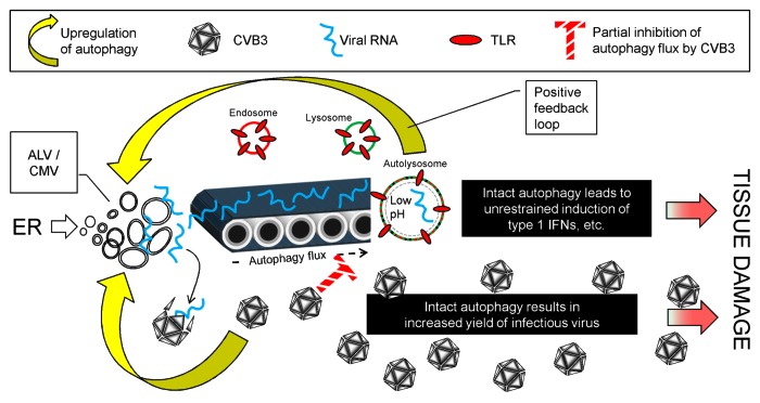
Figure 1. CVB3 and autophagy: Proposed interactions, and their pathogenic effects. CVB3 infection upregulates autophagy (lower yellow arrow), increasing the abundance of small autophagy-like vesicles (ALVs) which may provide a replication platform for the virus, allowing it to replicate to high titers, causing extensive disease (lower red arrow). Some of the viral RNA is captured by the autophagy pathway (conveyor belt) and ultimately encounters and activates TLRs in the autolysosome. This causes further upregulation of autophagy, thereby increasing the delivery of viral RNA to the TLRs; i.e., we propose that a positive feedback loop may exist (top yellow arrow). Viral proteins substantially, but incompletely, block autophagy flux (cross-hatched red T). These blocking effects may delay, but cannot prevent, the explosive innate response, which causes immunopathological damage, exacerbating disease (upper red arrow). In the absence of an intact autophagy pathway (Atg5f/f/Cre+ mice) both viral replication and TLR activation are reduced, thereby limiting disease.
Comment on
- Alirezaei M, Flynn CT, Wood MR, Whitton JL. Pancreatic acinar cell-specific autophagy disruption reduces coxsackievirus replication and pathogenesis in vivo. Cell Host Microbe. 2012;11:298–305. doi: 10.1016/j.chom.2012.01.014. doi: 10.1016/j.chom.2012.01.014
Publication types
MeSH terms
Grants and funding
LinkOut - more resources
Full Text Sources