. 2012 Jul 15;189(2):1002-13.
doi: 10.4049/jimmunol.1102624. Epub 2012 Jun 15.

Gemfibrozil, a lipid-lowering drug, upregulates IL-1 receptor antagonist in mouse cortical neurons: implications for neuronal self-defense

Affiliations

Gemfibrozil, a lipid-lowering drug, upregulates IL-1 receptor antagonist in mouse cortical neurons: implications for neuronal self-defense

Grant T Corbett et al. J Immunol. .

Abstract

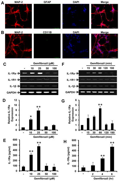
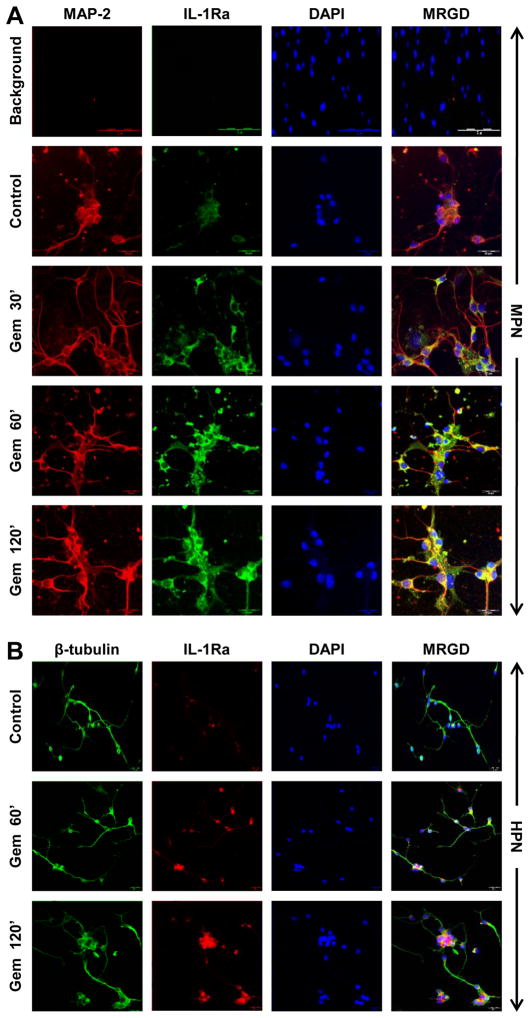
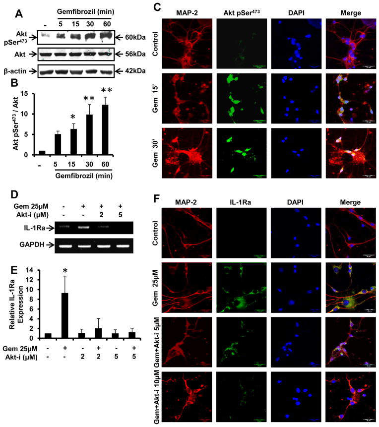
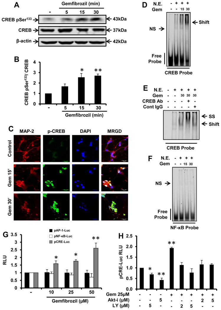
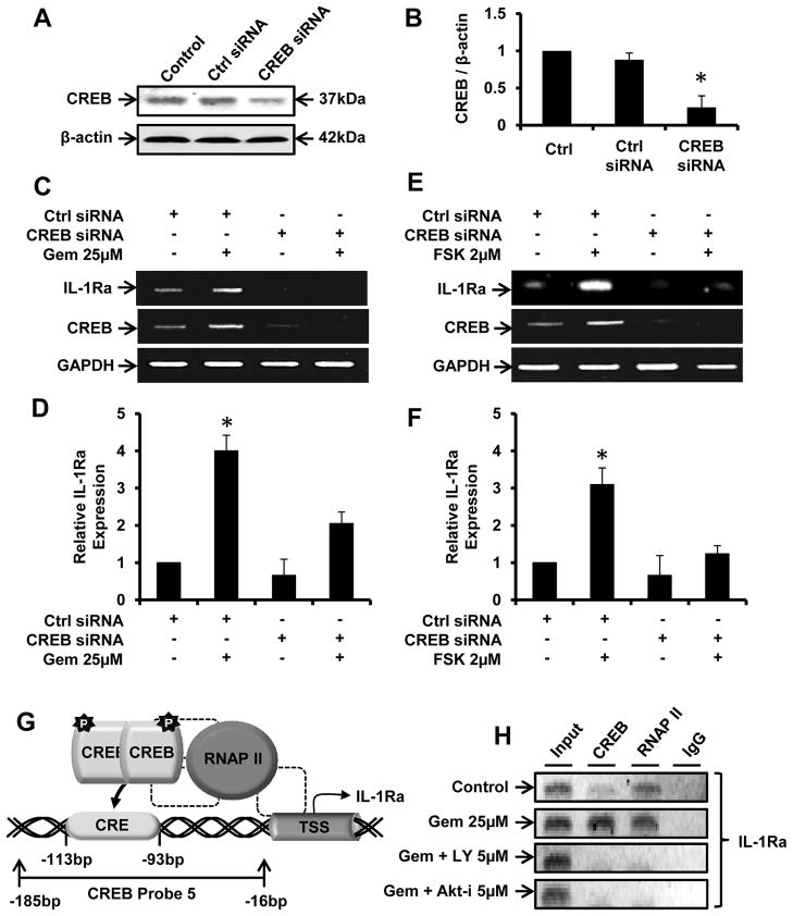
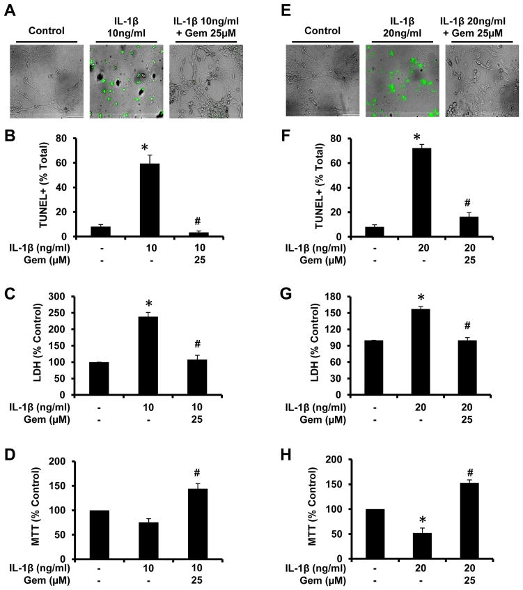
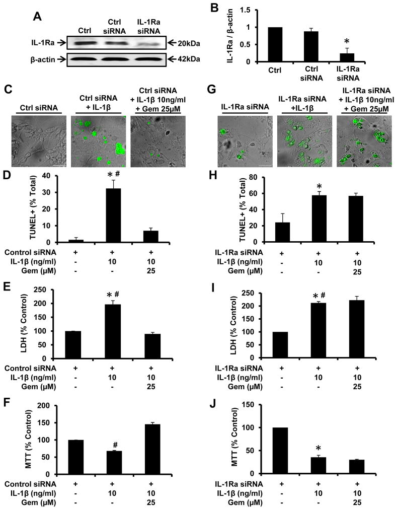
Chronic inflammation is becoming a hallmark of several neurodegenerative disorders and accordingly, IL-1β, a proinflammatory cytokine, is implicated in the pathogenesis of neurodegenerative diseases. Although IL-1β binds to its high-affinity receptor, IL-1R, and upregulates proinflammatory signaling pathways, IL-1R antagonist (IL-1Ra) adheres to the same receptor and inhibits proinflammatory cell signaling. Therefore, upregulation of IL-1Ra is considered important in attenuating inflammation. The present study underlines a novel application of gemfibrozil (gem), a Food and Drug Administration-approved lipid-lowering drug, in increasing the expression of IL-1Ra in primary mouse and human neurons. Gem alone induced an early and pronounced increase in the expression of IL-1Ra in primary mouse cortical neurons. Activation of type IA p110α PI3K and Akt by gem and abrogation of gem-induced upregulation of IL-1Ra by inhibitors of PI3K and Akt indicate a role of the PI3K-Akt pathway in the upregulation of IL-1Ra. Gem also induced the activation of CREB via the PI3K-Akt pathway, and small interfering RNA attenuation of CREB abolished the gem-mediated increase in IL-1Ra. Furthermore, gem was able to protect neurons from IL-1β insult. However, small interfering RNA knockdown of neuronal IL-1Ra abrogated the protective effect of gem against IL-1β, suggesting that this drug increases the defense mechanism of cortical neurons via upregulation of IL-1Ra. Taken together, these results highlight the importance of the PI3K-Akt-CREB pathway in mediating gem-induced upregulation of IL-1Ra in neurons and suggest gem as a possible therapeutic treatment for propagating neuronal self-defense in neuroinflammatory and neurodegenerative disorders.


Figures

Figure 1
Figure 1. Gemfibrozil upregulates the expression of IL-1Ra mRNA and protein in mouse cortical neurons
In order to determine the purity of neuronal cultures, chamber slides were double-labeled for MAP-2 and either GFAP (A) or CD11b (B). Fetal mouse cortical neurons (fMCNs) were treated with different concentrations of gemfibrozil for 1 hr followed by monitoring mRNA expression of IL-1Ra by semi-quantitative RT-PCR (C) and real-time PCR (D). fMCNs were treated with different concentrations of gemfibrozil for 6 hrs followed by measuring IL-1Ra protein in supernatants by sandwich ELISA (E). fMCNs were treated with 25μM gemfibrozil for 15 min, 30 min, 1 hr, 2 hr and 3 hr followed by monitoring the mRNA expression of IL-1Ra by semi quantitative RT-PCR (F) and real time PCR (G). fMCNs were treated with 25μM gemfibrozil for 2, 4 and 6 hrs followed by measuring IL-1Ra protein in supernatants by sandwich ELISA (H). Results are mean ± SD of three independent experiments. Significance is indicated by *p < 0.05 and **p < 0.01. Scale bar = 20μm.
Figure 2
Figure 2. Gemfibrozil upregulates IL-1Ra in fMCNs and human embryonic neurons
(A) fMCNs incubated with 25μM gemfibrozil for 30 min, 1 hr and 2 hrs were analyzed for IL-1Ra expression by immunofluorescence. Background fluorescence (A; top panel) and specificity (data not shown) of the IL-1Ra antibody was also monitored. (B) Primary human neurons treated with 25μM gemfibrozil for 1 and 2 hrs were analyzed for IL-1Ra expression by immunofluorescence. Scale bar = 20μm.
Figure 3
Figure 3. Gemfibrozil requires the activation of PI3-K to induce the upregulation of IL-1Ra in fMCNs
(A) Cells treated with 25μM gemfibrozil for 5, 15 and 30 min were analyzed for the presence of histone H3 and TrkB and recruitment of p110α, p110β and p110γ to the cellular membrane via western blot. (B) Densitometric analysis of time dependent recruitment of p110α to the plasma membrane. Cells preincubated with different concentrations of LY294002 and wortmannin for 30 min were treated with 25μM gemfibrozil for 1 hr followed by monitoring the expression of IL-1Ra mRNA by semi-quantitative RT-PCR (C) and real time PCR (D). The effects of LY294002 pretreatment on gemfibrozil-mediated induction of IL-1Ra protein were subsequently analyzed via immunofluorescence (E). Results are mean ± SD of three independent experiments. Significance is indicated by *p < 0.05 and **p < 0.01. Scale bar = 20μm.
Figure 4
Figure 4. Gemfibrozil induces IL-1Ra expression in fMCNs via activation of Akt
(A) Western blot analysis of phosphorylated Akt (p-Akt) and total Akt (t-Akt) in fMCNs treated with 25μM gemfibrozil for 5, 15, 30 and 60 min. (B) Densitometric analysis of time dependent increase in p-Akt by gemfibrozil. (C) Cells were treated with 25μM gemfibrozil for 15 and 30 min under the same culture conditions followed by monitoring the level of p-Akt by double-label immunofluorescence. Prior to treatment with gemfibrozil, neurons were preincubated with different concentrations of Akt-i for 30 min followed by monitoring the expression of IL-1Ra mRNA by semi-quantitative RT-PCR (D) and real time PCR (E) and protein by immunofluorescence (F). Results are mean ± SD of three independent experiments. Significance is indicated by *p < 0.05 and **p < 0.01. Scale bar = 20μm.
Figure 5
Figure 5. Gemfibrozil induces the activation of CREB in fMCNs via PI-3 kinase – Akt pathway
(A) Western blot analysis of phosphorylated CREB and total CREB in fMCNs treated with 25μM gemfibrozil for 5, 15 and 30 min. (B) Densitometric analysis of time dependent increase in phosphorylated CREB by gemfibrozil. (C) Cells were treated with 25μM gemfibrozil for 15 and 30 min followed by monitoring the level of phosphorylated CREB by double-label immunofluorescence. Cells were treated with 25μM gemfibrozil for 15 and 30 min followed by monitoring the DNA-binding activity of CREB (D) and NF-κB (F) by EMSA. (E) For supershift assay, nuclear extracts were incubated with 2 μg CREB antibody or control IgG for 30 min followed by EMSA. (G) Cells were transfected with different luciferase constructs (pNF-κB-Luc, pAP-1-Luc and pCRE-Luc) and 24 h after transfection, cells were stimulated with gemfibrozil. After 4 h, activity of firefly luciferase was measured in total cell extracts as described in Materials and Methods. (H) Cells were transfected with pCRE-Luc and after 24 h of transfection, cells were treated with different concentrations of LY294002 or Akt-i for 30 min followed by stimulation with gemfibrozil and luciferase assay. Results are mean ± SD of at least three independent experiments. Significance is indicated by *p < 0.05 and **p < 0.01. Scale bar = 20μm. N.E. = nuclear extract.
Figure 6
Figure 6. Involvement of CREB in gemfibrozil-induced transcription of IL-1Ra in fMCNs
Cells transfected with either control or CREB siRNA were analyzed for CREB inhibition efficiency via western blot (A) and subsequent densitometry (B). Forty-eight hours after transfection, cells were treated with either 25μM gemfibrozil (C & D) or 2μM forskolin (E & F) for 30 min followed by monitoring the mRNA expression of CREB and IL-1Ra by RT-PCR (C & E) and real time PCR (D & F). Results are mean ± SD of three independent experiments. Significance is indicated by *p < 0.05. (G) Graphical representation of the CRE located 93 to 113 base pairs upstream of the IL-1Ra transcription start site (TSS). (H) Cells preincubated with inhibitors of PI-3 kinase and Akt for 30 min were stimulated with gemfibrozil. After 2 h of stimulation, ChIP assay was performed as described in Materials and Methods.
Figure 7
Figure 7. Gemfibrozil protects fMCNs from IL-1β-induced apoptosis
Cells preincubated with 25μM gemfibrozil for 1 hr were exposed to either 10ng/ml (A, B, C, & D) or 20ng/ml (E, F, G, & H) IL-1β. After 2 hrs, cell death was monitored by phase-contrast TUNEL immunostaining (A & E), counting of TUNEL-positive cells (B & F), LDH release (C & G), and MTT (D & H). Results are mean ± SD of three independent experiments. Significance is indicated by *p < 0.05 relative to control and #p < 0.05 relative to the IL-1β. Scale bar = 20μm.
Figure 8
Figure 8. siRNA knockdown of IL-1Ra abrogates the protective efficacy of gemfibrozil from IL-1β-induced apoptosis in fMCNs
Cells were transfected with either control siRNA or IL-1Ra siRNA and monitored for IL-Ra inhibition efficiency via western blot (A) and subsequent densitometry (B). Cells transfected with either control siRNA (C, D, E & F) or IL-1Ra siRNA (G, H, I & J) were incubated with 25μM gemfibrozil for 1 hr followed by exposure to 10ng/ml IL-1β. After 2 hrs, cell death was monitored by phase-contrast TUNEL immunostaining (C & G), counting of TUNEL-positive cells (D & H), LDH release (E & I), and MTT (F & J). Results are mean ± SD of six independent experiments. Significance is indicated by *p < 0.05 relative to control and #p < 0.05 relative to the IL-1β. Scale bar = 20μm.

Similar articles

Cited by

References

    1. Alzheimer’s disease facts and figures. Alzheimer’s & dementia: the journal of the Alzheimer’s Association. 2011;7:208–244. - PubMed
    1. Jana A, Pahan K. Fibrillar amyloid-beta-activated human astroglia kill primary human neurons via neutral sphingomyelinase: implications for Alzheimer’s disease. J Neurosci. 30:12676–12689. - PMC - PubMed
    1. Martin JB. Molecular basis of the neurodegenerative disorders. N Engl J Med. 1999;340:1970–1980. - PubMed
    1. Cummings JL, Vinters HV, Cole GM, Khachaturian ZS. Alzheimer’s disease: etiologies, pathophysiology, cognitive reserve, and treatment opportunities. Neurology. 1998;51:S2–17. discussion S65–17. - PubMed
    1. Griffin WS, Sheng JG, Roberts GW, Mrak RE. Interleukin-1 expression in different plaque types in Alzheimer’s disease: significance in plaque evolution. J Neuropathol Exp Neurol. 1995;54:276–281. - PubMed

Publication types

MeSH terms

Substances