

Platelets present antigen in the context of MHC class I
- PMID: 22706078
- PMCID: PMC3392496
- DOI: 10.4049/jimmunol.1200580
Platelets present antigen in the context of MHC class I
Abstract
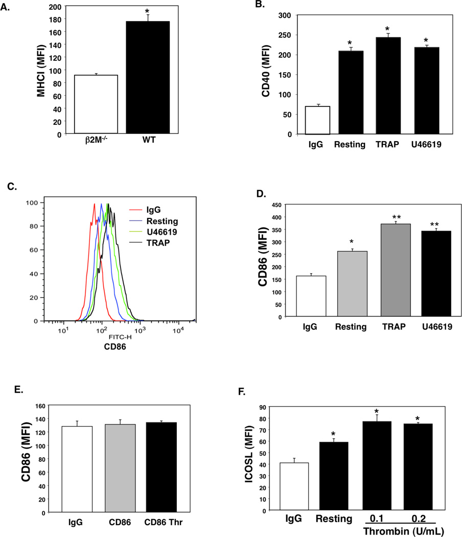
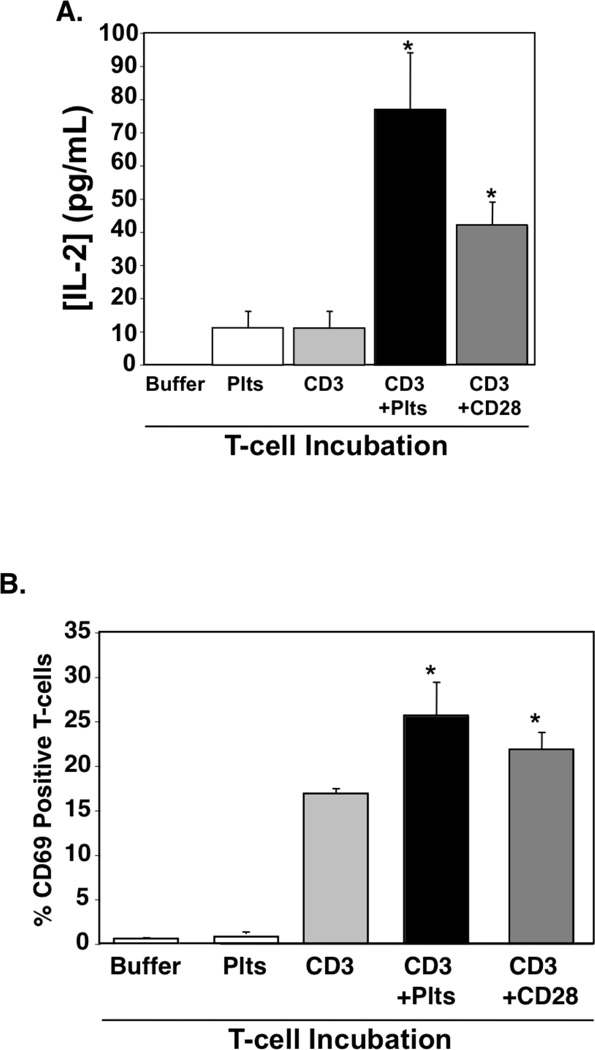
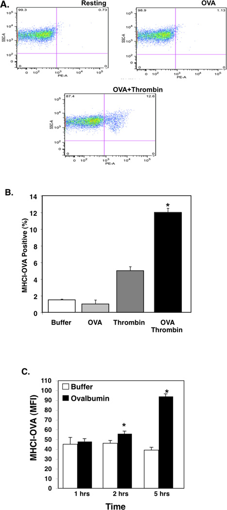
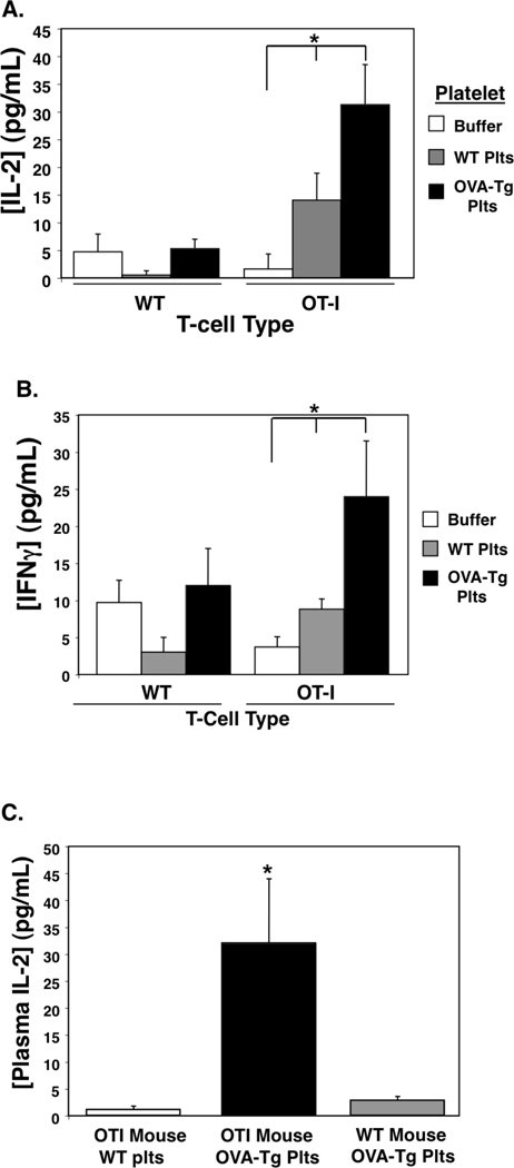
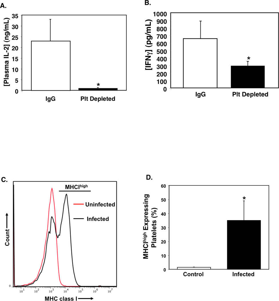
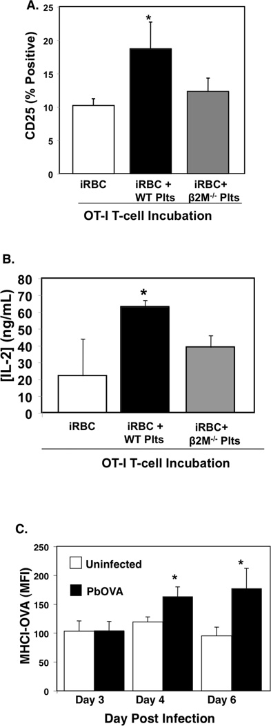
Platelets are most recognized for their vital role as the cellular mediator of thrombosis, but platelets also have important immune functions. Platelets initiate and sustain vascular inflammation in many disease conditions, including arthritis, atherosclerosis, transplant rejection, and severe malaria. We now demonstrate that platelets express T cell costimulatory molecules, process and present Ag in MHC class I, and directly activate naive T cells in a platelet MHC class I-dependent manner. Using an experimental cerebral malaria mouse model, we also demonstrate that platelets present pathogen-derived Ag to promote T cell responses in vivo, and that platelets can be used in a cell-based vaccine model to induce protective immune responses. Our study demonstrates a novel Ag presentation role for platelets.
Conflict of interest statement
The authors have not financial conflicts of interest.
Figures

Similar articles
-
MHC class I-dependent presentation of exoerythrocytic antigens to CD8+ T lymphocytes is required for protective immunity against Plasmodium berghei.J Immunol. 1996 May 1;156(9):3374-81. J Immunol. 1996. PMID: 8617963
-
TAP-mediated processing of exoerythrocytic antigens is essential for protection induced with radiation-attenuated Plasmodium sporozoites.Eur J Immunol. 2016 Apr;46(4):885-96. doi: 10.1002/eji.201545748. Epub 2016 Jan 21. Eur J Immunol. 2016. PMID: 26703789
-
Direct processing and presentation of antigen from malaria sporozoites by professional antigen-presenting cells in the induction of CD8 T-cell responses.Immunol Cell Biol. 2005 Jun;83(3):307-12. doi: 10.1111/j.1440-1711.2005.01325.x. Immunol Cell Biol. 2005. PMID: 15877610
-
Comparative study of the role of professional versus semiprofessional or nonprofessional antigen presenting cells in the rejection of vascularized organ allografts.Transpl Immunol. 1995 Dec;3(4):273-89. doi: 10.1016/0966-3274(95)80013-1. Transpl Immunol. 1995. PMID: 8665146 Review.
-
Role of autophagy in MHC class I-restricted antigen presentation.Mol Immunol. 2019 Sep;113:2-5. doi: 10.1016/j.molimm.2017.10.021. Epub 2017 Nov 8. Mol Immunol. 2019. PMID: 29126597 Free PMC article. Review.
Cited by
-
Platelet-derived β2m regulates age related monocyte/macrophage functions.Aging (Albany NY). 2019 Dec 18;11(24):11955-11974. doi: 10.18632/aging.102520. Epub 2019 Dec 18. Aging (Albany NY). 2019. PMID: 31852838 Free PMC article.
-
Pneumonitis after immune checkpoint inhibitor therapies in patients with acute myeloid leukemia: A retrospective cohort study.Cancer. 2022 Jul 15;128(14):2736-2745. doi: 10.1002/cncr.34229. Epub 2022 Apr 22. Cancer. 2022. PMID: 35452134 Free PMC article.
-
Platelet-lymphocyte co-culture serves as an ex vivo platform of dynamic heterotypic cross-talk.J Cell Commun Signal. 2022 Dec;16(4):661-675. doi: 10.1007/s12079-022-00676-0. Epub 2022 Apr 12. J Cell Commun Signal. 2022. PMID: 35414144 Free PMC article.
-
Platelets in pediatric and neonatal sepsis: novel mediators of the inflammatory cascade.Pediatr Res. 2022 Jan;91(2):359-367. doi: 10.1038/s41390-021-01715-z. Epub 2021 Oct 28. Pediatr Res. 2022. PMID: 34711945 Free PMC article. Review.
-
The Non-Hemostatic Aspects of Transfused Platelets.Front Med (Lausanne). 2018 Feb 27;5:42. doi: 10.3389/fmed.2018.00042. eCollection 2018. Front Med (Lausanne). 2018. PMID: 29536007 Free PMC article. Review.
References
-
- Massberg S, Konrad I, Schurzinger K, Lorenz M, Schneider S, Zohlnhoefer D, Hoppe K, Schiemann M, Kennerknecht E, Sauer S, Schulz C, Kerstan S, Rudelius M, Seidl S, Sorge F, Langer H, Peluso M, Goyal P, Vestweber D, Emambokus NR, Busch DH, Frampton J, Gawaz M. Platelets secrete stromal cell-derived factor 1alpha and recruit bone marrow-derived progenitor cells to arterial thrombi in vivo. J Exp Med. 2006;203:1221–1233. - PMC - PubMed
-
- Gawaz M. Role of platelets in coronary thrombosis and reperfusion of ischemic myocardium. Cardiovasc Res. 2004;61:498–511. - PubMed
-
- Langer H, May AE, Daub K, Heinzmann U, Lang P, Schumm M, Vestweber D, Massberg S, Schonberger T, Pfisterer I, Hatzopoulos AK, Gawaz M. Adherent platelets recruit and induce differentiation of murine embryonic endothelial progenitor cells to mature endothelial cells in vitro. Circ Res. 2006;98:e2–e10. - PubMed
-
- Khandoga A, Hanschen M, Kessler JS, Krombach F. CD4+ T cells contribute to postischemic liver injury in mice by interacting with sinusoidal endothelium and platelets. Hepatology. 2006;43:306–315. - PubMed
Publication types
MeSH terms
Substances
Grants and funding
LinkOut - more resources
Full Text Sources
Other Literature Sources
Molecular Biology Databases
Research Materials
