

Cutting edge: mast cells critically augment myeloid-derived suppressor cell activity
- PMID: 22706087
- PMCID: PMC3392490
- DOI: 10.4049/jimmunol.1200647
Cutting edge: mast cells critically augment myeloid-derived suppressor cell activity
Abstract
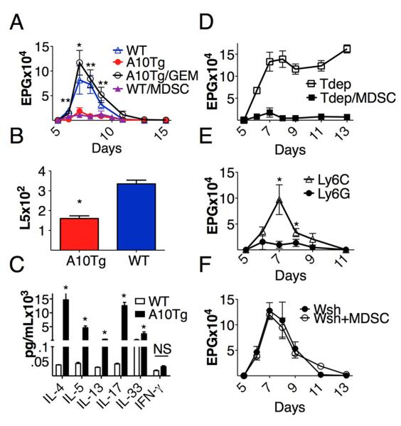
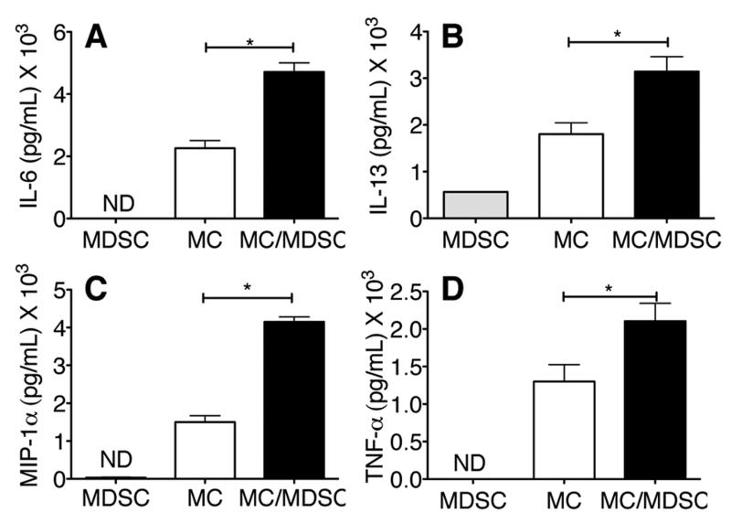
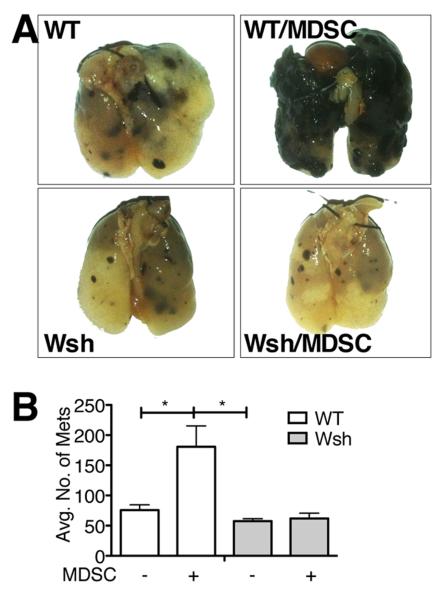
Myeloid-derived suppressor cells (MDSCs) are primarily recognized for their immunosuppressive properties in malignant disease. However, their interaction with other innate immune cells and their regulation of immune responses, such as in parasitic infection, necessitate further characterization. We used our previously published mouse model of MDSC accumulation to examine the immunoregulatory role of MDSCs in B16 melanoma metastasis and Nippostrongylus brasiliensis infection. In this study, we demonstrate that the activity of MDSCs is dependent on the immune stimuli and subset induced. Monocytic MDSCs predictably suppressed antitumor immune responses but granulocytic MDSCs surprisingly enhanced the clearance of N. brasiliensis infection. Intriguingly, both results were dependent on MDSC interaction with mast cells (MCs), as demonstrated by adoptive-transfer studies in MC-deficient (Kit(Wsh)(/)(Wsh)) mice. These findings were further supported by ex vivo cocultures of MCs and MDSCs, indicating a synergistic increase in cytokine production. Thus, MCs can enhance both immunosuppressive and immunosupportive functions of MDSCs.
Figures

Similar articles
-
Myeloid-derived suppressor cells enhance IgE-mediated mast cell responses.J Leukoc Biol. 2014 Apr;95(4):643-50. doi: 10.1189/jlb.0913510. Epub 2013 Dec 12. J Leukoc Biol. 2014. PMID: 24338630 Free PMC article.
-
Mast cell histamine promotes the immunoregulatory activity of myeloid-derived suppressor cells.J Leukoc Biol. 2014 Jul;96(1):151-9. doi: 10.1189/jlb.5A1213-644R. Epub 2014 Mar 7. J Leukoc Biol. 2014. PMID: 24610880 Free PMC article.
-
Tumor-infiltrating monocytic myeloid-derived suppressor cells mediate CCR5-dependent recruitment of regulatory T cells favoring tumor growth.J Immunol. 2012 Dec 15;189(12):5602-11. doi: 10.4049/jimmunol.1201018. Epub 2012 Nov 14. J Immunol. 2012. PMID: 23152559
-
On the origin of myeloid-derived suppressor cells.Oncotarget. 2017 Jan 10;8(2):3649-3665. doi: 10.18632/oncotarget.12278. Oncotarget. 2017. PMID: 27690299 Free PMC article. Review.
-
Reciprocal relationship between myeloid-derived suppressor cells and T cells.J Immunol. 2013 Jul 1;191(1):17-23. doi: 10.4049/jimmunol.1300654. J Immunol. 2013. PMID: 23794702 Free PMC article. Review.
Cited by
-
Mast Cells: A New Frontier for Cancer Immunotherapy.Cells. 2021 May 21;10(6):1270. doi: 10.3390/cells10061270. Cells. 2021. PMID: 34063789 Free PMC article. Review.
-
Tolerance and immune suppression in the tumor microenvironment.Cell Immunol. 2016 Jan;299:23-9. doi: 10.1016/j.cellimm.2015.09.011. Epub 2015 Sep 30. Cell Immunol. 2016. PMID: 26435343 Free PMC article.
-
Here, There, and Everywhere: Myeloid-Derived Suppressor Cells in Immunology.J Immunol. 2023 May 1;210(9):1183-1197. doi: 10.4049/jimmunol.2200914. J Immunol. 2023. PMID: 37068300 Free PMC article. Review.
-
Frenemies in the Microenvironment: Harnessing Mast Cells for Cancer Immunotherapy.Pharmaceutics. 2023 Jun 9;15(6):1692. doi: 10.3390/pharmaceutics15061692. Pharmaceutics. 2023. PMID: 37376140 Free PMC article. Review.
-
Translocation of Helicobacter hepaticus synergizes with myeloid-derived suppressor cells and contributes to breast carcinogenesis.Oncoimmunology. 2022 Mar 26;11(1):2057399. doi: 10.1080/2162402X.2022.2057399. eCollection 2022. Oncoimmunology. 2022. PMID: 35371619 Free PMC article.
References
-
- Narita Y, Wakita D, Ohkur T, Chamoto K, Nishimura T. Potential differentiation of tumor bearing mouse CD11b+Gr-1+ immature myeloid cells into both suppressor macrophages and immunostimulatory dendritic cells. Biomed. Res. 2009;30:7–15. - PubMed
Publication types
MeSH terms
Grants and funding
LinkOut - more resources
Full Text Sources
Other Literature Sources
Molecular Biology Databases
