. 2012 Jun 17;15(7):979-87.
doi: 10.1038/nn.3135.

Distinct molecular pathways mediate glial activation and engulfment of axonal debris after axotomy

Affiliations

Distinct molecular pathways mediate glial activation and engulfment of axonal debris after axotomy

Jennifer S Ziegenfuss et al. Nat Neurosci. .

Abstract

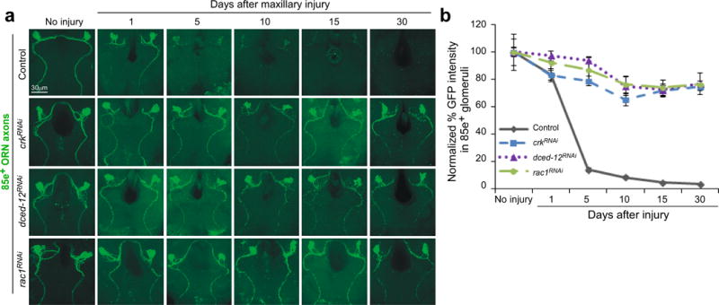
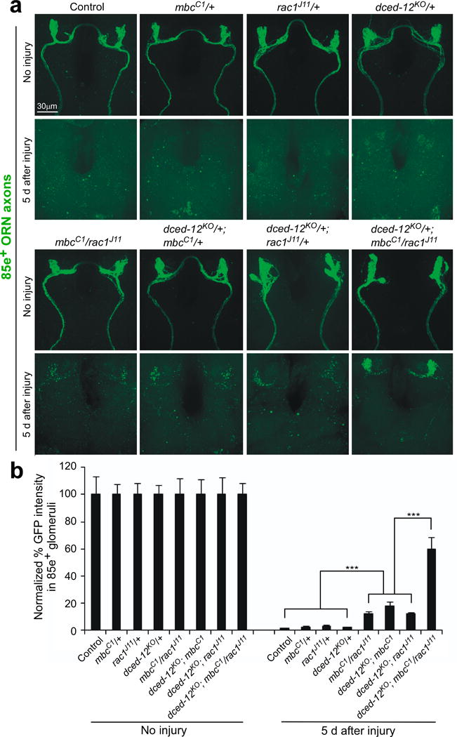
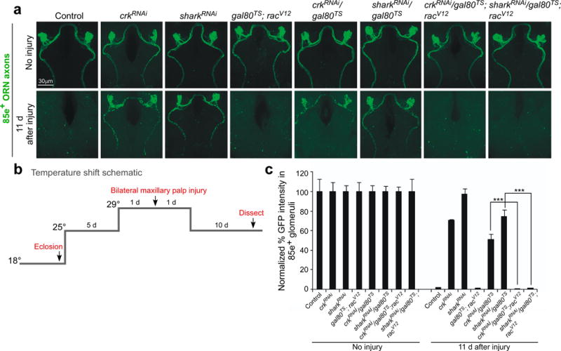
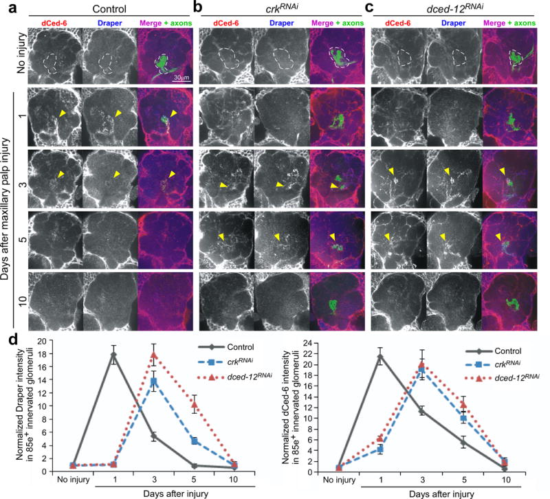
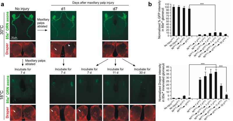
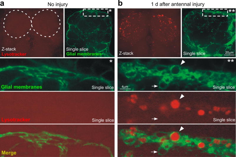
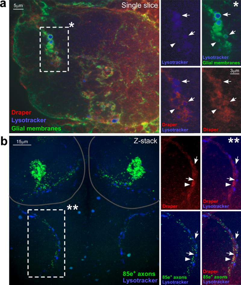
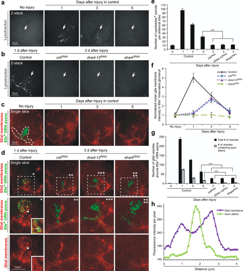
Glial cells efficiently recognize and clear cellular debris after nervous system injury to maintain brain homeostasis, but pathways governing glial responses to neural injury remain poorly defined. We identify the Drosophila melanogaster guanine nucleotide exchange factor complex Crk/Mbc/dCed-12 and the small GTPase Rac1 as modulators of glial clearance of axonal debris. We found that Crk/Mbc/dCed-12 and Rac1 functioned in a non-redundant fashion with the Draper transmembrane receptor pathway: loss of either pathway fully suppressed clearance of axonal debris. Draper signaling was required early during glial responses, promoting glial activation, which included increased Draper and dCed-6 expression and extension of glial membranes to degenerating axons. In contrast, the Crk/Mbc/dCed-12 complex functioned at later phases, promoting glial phagocytosis of axonal debris. Our work identifies new components of the glial engulfment machinery and shows that glial activation, phagocytosis of axonal debris and termination of responses to injury are genetically separable events mediated by distinct signaling pathways.


Figures

Figure 1
Figure 1. Drosophila Crk, dCed-12, and Rac1 are required for glial clearance of severed axons from the CNS
(a) OR85e-expressing ORNs were labeled with mCD8::GFP in control (w;OR85e-mCD8::GFP/+; repo-Gal4/+) and glial RNAi for crkRNAi (w;OR85e-mCD8::GFP/UAS-crk19061RNAi; repo-Gal4/+), dced-12RNAi (w;OR85e-mCD8::GFP/+; repo-Gal4/UAS-dced-1210455RNAi), and rac1RNAi (w, UAS-rac149247RNAi/+ ;OR85e-mCD8::GFP/+; repo-Gal4/+). Maxillary palps were bilaterally ablated and clearance of axonal debris from the CNS was assayed with anti-GFP antibody stains (green) 1, 5, 10, 15, and 30 days after injury. (b) Normalized quantification of data to control from (a) Error bars represent s.e.m.; n>10 for all.
Figure 2
Figure 2. mbc, dced-12, and rac1 mutants exhibit dominant genetic interactions in ORN axon clearance assays
(a) OR85e-expressing ORN axons were labeled in control (w;OR85e-mCD8::GFP/+) animals and in mbcC1, dced-12KO, or rac1J11 heterozygous mutant backgrounds, maxillary palps were ablated, or left uninjured, and clearance of axons was assayed after 5 days (anti-GFP, green). (b) Normalized quantification of data to uninjured cohorts from (a) Error bars represent s.e.m.; n>10; *** p<0.0001.
Figure 3
Figure 3. Constitutively active Rac rescues both Draper signaling and GEF pathway engulfment defects
(a) OR85e-expressing ORNs were labeled with mCD8::GFP in control (w;OR85e-mCD8::GFP/+;TIFR-Gal4/+) and glial RNAi for crk (w;OR85e-mCD8::GFP, UAS-crk106498RNAi /+;TIFR-Gal4/+) and shark (w;OR85e-mCD8::GFP, UAS-shark6bRNAi /+;TIFR-Gal4/+) either containing gal80TS (w;OR85e-mCD8::GFP; UAS-crk106498RNAi /gal80TS;TIFR-Gal4/+ and w;OR85e-mCD8::GFP, UAS-shark6bRNAi /gal80TS;TIFR-Gal4/+) or with constitutive active racV12 (w;OR85e-mCD8::GFP; UAS-crk106498RNAi /gal80TS;TIFR-Gal4/racV12 and w;OR85e-mCD8::GFP, UAs-shark6bRNAi /gal80TS;TIFR-Gal4/racV12 and w;OR85e-mCD8::GFP/gal80TS;racV12/+). Maxillary palps were bilaterally ablated and clearance of axonal debris was assayed with anti-GFP antibody stains (green) 11 days after injury, according to the temperature shift protocol outlined in (b). (c) Normalized quantification of data to uninjured cohorts from (a) Error bars represent s.e.m.; n>10; *** p<0.0001.
Figure 4
Figure 4. Crk and dCed-12 are not required for activation of glia after axotomy
(a, b, and c) OR85e-expressing ORNs were labeled with mCD8::GFP in control (w;OR85e-mCD8::GFP/+; repo-Gal4/+) and those with glia-specific knockdown of crk and dced-12 (w;UAS-crk19061RNAi/ OR85e-mCD8::GFP;repo-Gal4/+ and w; OR85e-mCD8::GFP/+; repo-Gal4/UAS-dced-1210455RNAi). Animals were assayed for injury-induced recruitment of dCed-6 (red, pseudo-colored grey, left panel) and Draper (blue, pseudo-colored grey middle panel) to severed axons in maxillary-palp innervated glomeruli (arrows) 1, 3, 5, and 10 days after injury. (d) Normalized quantification to uninjured cohorts of Draper and dCed-6 intensity in 85e-innervated glomeruli (right panel, dotted outline) from (a, b, and c). Error bars represent s.e.m.; n>10 for all.
Figure 5
Figure 5. Axons remain viable engulfment targets capable of activating glia for over a week after axotomy
(a) Experimental animals (w;OR85e-mCD8::GFP/UAS-shibirets; mz0709-Gal4/+) were raised at the permissive temperature, 18°C, and kept at 18°C for 7 days after eclosion. Adult animals were then shifted to the restrictive temperature, 30°C, and either left uninjured or injured via bilateral maxillary palp ablation. Left, expression of Shibirets in ensheathing glia where animals were returned to 18°C, injured, and allowed 7 days to clear axonal debris. Cohorts kept at 30°C (where glial engulfment activity was “frozen”) were assayed for changes in glial Draper expression (red) around maxillary-innervated glomeruli (arrows) and clearance of axonal debris (anti-GFP, green) at 1 (D1) or 7 (D7) days after injury at 30°C. Arrowhead; appearance of Draper+ puncta only at periphery of antennal lobe. These same cohorts were returned to 18°C and allowed 7, 11, or 30 additional days to clear degenerating axons. (b) Normalized quantification of data to cohort controls from (a) Error bars represent s.e.m.; n>10; ***, P<0.0001.
Figure 6
Figure 6. Axotomy-induced activation of phagolysosomes in engulfing glia
(a) Uninjured animals (w;UAS-mcd8::GFP/+;TIFR-Gal4/+) were stained for Lysotracker Red (confocal Z-stack, top) and visualized for GFP+ glial membranes (single confocal slice, top). Dotted circles; location of antennal lobes. *; high magnification view of the marked rectangle in a single confocal section. (b) Animals (w;UAS-mcd8::GFP/+;TIFR-Gal4/+) shown 1 day after bilateral antennal injury were stained for Lysotracker Red (Z-stack top) and visualized for GFP+ glial membranes (single slice, top). **; high magnification view of the marked rectangle in a single confocal section. Arrow; an injury-induced glial vesicle. Arrowhead; glial vesicle positive for lysosomal activity.
Figure 7
Figure 7. Glial phagolysosomes are decorated with Draper and preferentially accumulate around degenerating axons
(a) Animals (w;UAS-mcd8::GFP/+;TIFR-Gal4/+) shown 1 day after bilateral maxillary palp injury were stained for Draper (pseudo-colored red), Lysotracker Red (pseudo-colored blue), and visualized for GFP+ glial membranes. *; high magnification view of the marked rectangle in a single confocal section. Arrows; injury-induced vesicles positive for Draper recruitment and lysosomal activity. Arrowhead; vesicles positive for Draper recruitment but lacking lysosomal activity. (b) Animals (w;OR85e-mCD8::GFP/+) shown 1 day after bilateral maxillary palp injury were stained for GFP (green), Draper (pseudo-colored red) and Lysotracker Red (pseudo-colored blue). Circles; location of antennal lobes. **; high magnification view of the marked rectangle in a confocal Z-stack. Arrows; areas of lysosomal activity with little to no remaining GFP+ axon debris. Arrowhead; areas where groups of axon debris co-localize with Draper along the degenerating maxillary palp nerve.
Figure 8
Figure 8. Crk and dCed-12 are required for internalization of axonal debris and phagolysosome formation
(a) Lysosomal activity was monitored with Lysotracker Red (pseudo-colored grey) in control (w; TIFR-Gal4/+) animals in individual antennal lobes (pictured) 1, 3, and 5 days after maxillary palp injury. Arrows; location of 85e-innervated maxillary-palp glomeruli. Representative single confocal Z-sections are shown for all. (b) Lysosomal activity was monitored in control (w; TIFR-Gal4/+) animals 1 day after maxillary palp injury, and 3 days after injury in glial RNAi animals for crk106498RNAi, dced-1210455RNAi, and shark6bRNAi. Arrows; location of 85e-innervated maxillary-palp glomeruli. (c) Glial membranes were labeled with tdTomato, OR85e-expressing axons (anti-GFP, green) were labeled with mCD8::GFP (w;OR85e-mCD8::GFP/UAS-cd4-tdTomato;TIFR-Gal4/+) and we monitored glial membrane recruitment to axonal debris, and the formation of vesicles within ensheathing glia 0, 1, 3, and 5 days after maxillary palp injury. Outlined; 85e-innervated maxillary-palp glomerulus used to quantify glial invasion and vesicle formation into injured area. (d) Glial membranes and axons were labeled as in (d). Recruitment of glial membranes to degenerating axons, and internalization of axonal debris by ensheathing glia was scored in control and glial crk106498RNAi, dced-1210455RNAi,and shark6bRNAi animals. (a’-d’); high magnification views of marked rectangle areas. Arrowhead; red glial vesicle containing axon debris. Lower right corner box in a’: high magnification view of debris-containing glial vesicles. (e) Quantification of antennal lobe lysosomal puncta data from (a, b).; Error bars represent s.e.m.; n>10; ***, P<0.0001. (f) Normalized quantification to control from (d, e) of glial membrane infiltration around degenerating 85e+ axons. (g) Quantification from (d, e) of the total number of glial vesicles and number of vesicles containing axonal debris around 85e-innervated maxillary-palp glomeruli.; Error bars represent s.e.m.; n>10; ***, P<0.0001. (h) Example from corner box in (a’) of fluorescence intensity of the glial membrane and axon debris along line drawn through an individual glial vesicle.

Similar articles

Cited by

References

    1. Cuadros MA, Navascues J. The origin and differentiation of microglial cells during development. Prog Neurobiol. 1998;56:173–189. - PubMed
    1. Marin-Teva JL, Cuadros MA, Calvente R, Almendros A, Navascues J. Naturally occurring cell death and migration of microglial precursors in the quail retina during normal development. J Comp Neurol. 1999;412:255–275. - PubMed
    1. Parnaik R, Raff MC, Scholes J. Differences between the clearance of apoptotic cells by professional and non-professional phagocytes. Curr Biol. 2000;10:857–860. - PubMed
    1. Block ML, Zecca L, Hong JS. Microglia-mediated neurotoxicity: uncovering the molecular mechanisms. Nat Rev Neurosci. 2007;8:57–69. - PubMed
    1. Sofroniew MV. Molecular dissection of reactive astrogliosis and glial scar formation. Trends Neurosci. 2009;32:638–647. - PMC - PubMed

Publication types

LinkOut - more resources