

Primary biliary tract melanoma: Report of a case and review of the literature
- PMID: 22706296
- PMCID: PMC3397286
- DOI: 10.1016/j.ijscr.2012.05.008
Primary biliary tract melanoma: Report of a case and review of the literature
Abstract
Introduction: Primary melanoma of the bile duct is extremely rare with only nine cases of primary melanoma of the bile duct reported in the literature.
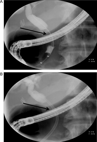
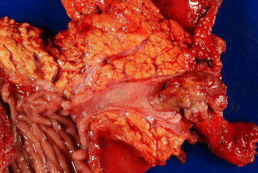
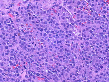
Presentation of case: A 55-year-old previously healthy gentleman developed increasing jaundice over several months and subsequently underwent an ERCP with stone extraction. Cytology brushings in an area of a distal stricture in the bile duct were concerning for cholangiocarcinoma. The patient was referred to our institution and underwent a pancreaticoduodenectomy. The surgical specimen showed a single 4.5cm polypoid lesion located in the bile duct. A diagnosis of melanoma was rendered after immunohistochemical studies on the tumor demonstrated positivity for melanoma markers. Follow-up of the patient with skin, ocular, and lymph node exams showed no evidence of melanoma. A PET scan 4 and 10 months post-surgery failed to reveal either a primary skin lesion or other sites of metastases.
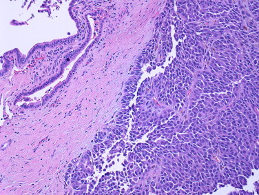
Discussion: The vast majority of melanomas of the bile duct represent metastases from a cutaneous source and tend to present as multiple flat pigmented lesions. Conversely, cases of primary bile duct melanoma are characterized by a distinct gross morphology consisting of a solitary intraluminal polypoid lesion attached by a pedicle with no other identifiable primary lesion. Other supporting criteria include absence of other involved sites and presence of an in situ junctional component.
Conclusion: Given the clinical history, gross findings, and lack of a primary cutaneous site or other demonstrable metastases, this patient likely represents the tenth reported case of primary biliary tract melanoma.
Copyright © 2012 Surgical Associates Ltd. Published by Elsevier Ltd. All rights reserved.
Figures
References
-
- Martel J.P., McLean C.A., Rankin R.N. Melanoma of the gallbladder. Radiographics. 2009;29(January–February (1)):291–296. - PubMed
-
- Zaide E. Malignant melanoma of the choledochus. Arquivos de Oncología. 1963;26:254–255. - PubMed
-
- Carstens H.B., Ghazi C., Carnighan R.H., Brewer M.S. Primary malignant melanoma of the common bile duct. Human Pathology. 1986;17(December (12)):1282–1285. - PubMed
-
- Wright R.A., Brewer M. Primary melanoma of the common bile duct. Southern Medical Journal. 1988;81(March (3)):396–397. - PubMed
-
- Deugnier Y., Turlin B., Lehry D., Pennarun J.R., Verger P., Launois B. Malignant melanoma of the hepatic and common bile ducts. A case report and review of the literature. Archives of Pathology and Laboratory Medicine. 1991;115(September (9)):915–917. - PubMed
LinkOut - more resources
Full Text Sources
Research Materials
