. 2012 Jul;122(7):2482-98.
doi: 10.1172/JCI63239. Epub 2012 Jun 18.

Fungal antioxidant pathways promote survival against neutrophils during infection

Affiliations

Fungal antioxidant pathways promote survival against neutrophils during infection

Sixto M Leal Jr et al. J Clin Invest. 2012 Jul.

Abstract

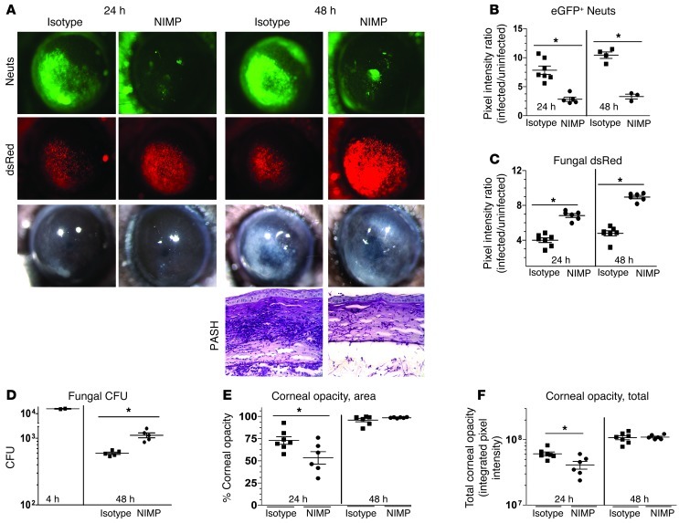
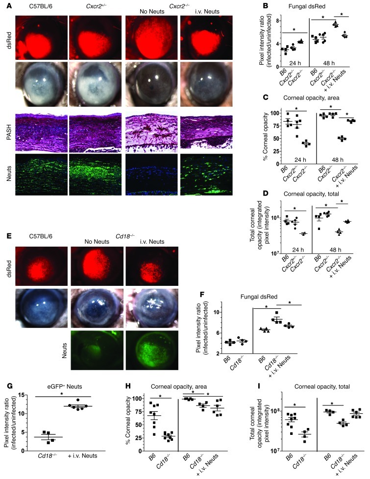
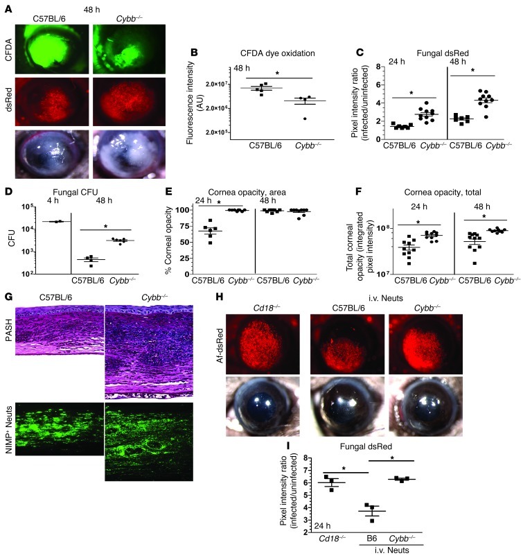
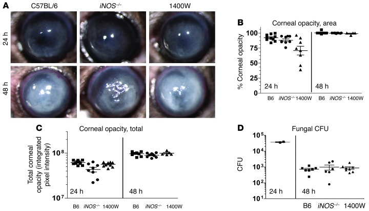
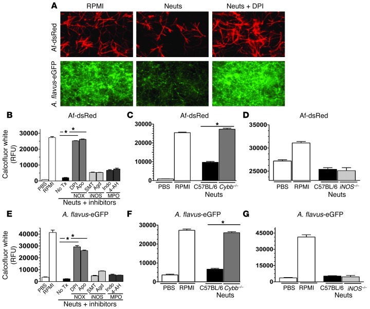
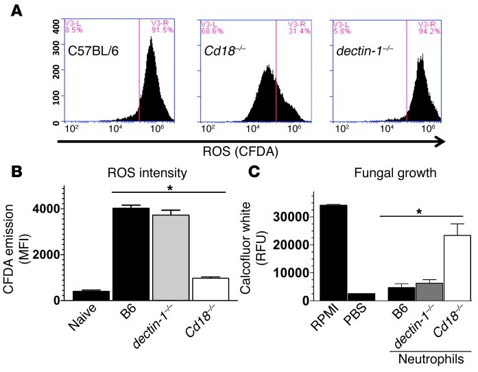
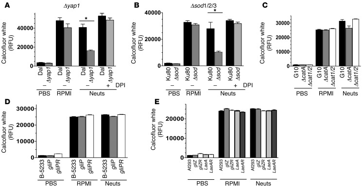
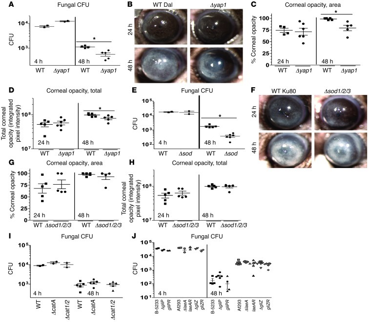
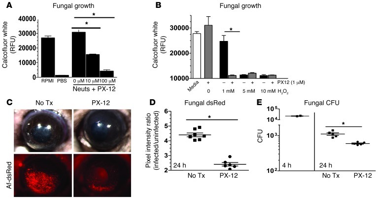
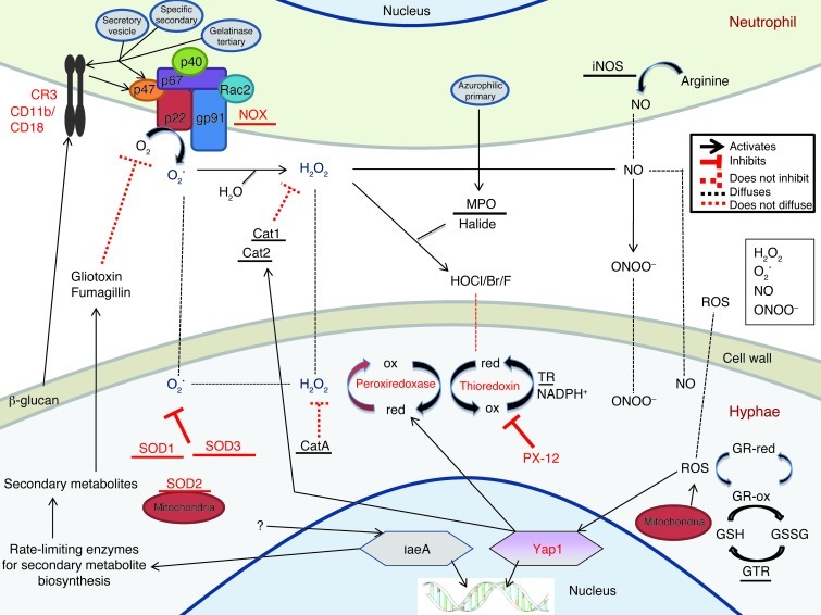
Filamentous fungi are a common cause of blindness and visual impairment worldwide. Using both murine model systems and in vitro human neutrophils, we found that NADPH oxidase produced by neutrophils was essential to control the growth of Aspergillus and Fusarium fungi in the cornea. We demonstrated that neutrophil oxidant production and antifungal activity are dependent on CD18, but not on the β-glucan receptor dectin-1. We used mutant A. fumigatus strains to show that the reactive oxygen species-sensing transcription factor Yap1, superoxide dismutases, and the Yap1-regulated thioredoxin antioxidant pathway are each required for protection against neutrophil-mediated oxidation of hyphae as well as optimal survival of fungal hyphae in vivo. We also demonstrated that thioredoxin inhibition using the anticancer drug PX-12 increased the sensitivity of fungal hyphae to both H2O2- and neutrophil-mediated killing in vitro. Additionally, topical application of PX-12 significantly enhanced neutrophil-mediated fungal killing in infected mouse corneas. Cumulatively, our data reveal critical host oxidative and fungal anti-oxidative mediators that regulate hyphal survival during infection. Further, these findings also indicate that targeting fungal anti-oxidative defenses via PX-12 may represent an efficacious strategy for treating fungal infections.


Figures

Figure 1
Figure 1. Neutrophil depletion enhances fungal growth during corneal infection.
(A) Transgenic C57BL/6 mice with neutrophil-specific eGFP expression downstream of the lysozyme promotor (LysM) were depleted of neutrophils (Neuts) with neutrophil-specific NIMPR-14 antibody (i.p.) and infected with 40,000 Af-dsRed conidia. Eyes were imaged at 24 and 48 hours after infection for neutrophil infiltration (eGFP), fungal growth (dsRed), and corneal opacity (BF). In addition, PASH stains were performed on 5-μm sections of corneas at 48 hours after infection. (B) MetaMorph software was used to quantify neutrophil infiltration (eGFP emission) and (C) fungal dsRed expression. (D) At 4 and 48 hours after infection, eyes were homogenized and plated on SDA plates and CFU quantified by direct counting. MetaMorph software was utilized to quantify (E) corneal opacity area and (F) total corneal opacity (described in detail in Supplemental Figure 1). Three independent experiments (n = 5) were performed. *P < 0.05. Original magnification, ×20 (eye images); ×400 (histology).
Figure 2
Figure 2. Neutrophil adoptive transfer restricts fungal growth during corneal infection.
(A) C57BL/6, Cxcr2+/–, and Cxcr2–/– mice were infected with 30,000 Af-dsRed conidia. At 24 hours after infection, one group of fungus-infected Cxcr2–/– mice were injected i.v. with 4 × 10⁶ BMNs from C57BL/6 (B6) mice. At 24 hours after infection, corneas were imaged for cellular infiltration, fungal growth, and corneal opacity. At this time point, mice were euthanized, eyes were fixed in formalin, and 5-μm corneal sections were PASH stained. (B) MetaMorph software was used to quantify fungal dsRed expression, (C) corneal opacity area, and (D) total corneal opacity in infected corneas. (E) Similar to Cxcr2–/– mice, C57BL/6 and Cd18–/– mice were infected with Af-dsRed conidia, and at 24 hours after infection one group of infected Cd18–/– mice were given 4 million adoptively transferred BMNs isolated from a LysM-eGFP mouse (eGFP+ Neuts) and eyes were imaged at 48 hours. (F) Fungal dsRed expression, (G) eGFP+ neutrophil infiltration, (H) corneal opacity area, and (I) total corneal opacity were quantified using MetaMorph software. Three independent experiments (n = 5) were performed. *P < 0.05. Original magnification, ×20 (eye images); ×400 (histology).
Figure 3
Figure 3. Neutrophil NOX is required for control of A. fumigatus fungal growth during corneal infection.
(A) C57BL/6 mice and Cybb–/– mice were infected with 40,000 A. fumigatus strain Af-dsRed conidia. Eyes were imaged at 48 hours after infection for ROS-mediated CFDA dye oxidation, fungal dsRed expression, and corneal opacity. (B) CFDA dye oxidation, (C) fungal dsRed expression, (D) CFU, (E) corneal opacity area, and (F) total corneal opacity were quantified after infection. (G) 5-μm sections of 48-hour-infected fungal corneas were stained with PASH or neutrophil-specific NIMP antibody. (H) Cd18–/– mice were infected with A. fumigatus strain Af-dsRed conidia. At 2 hours after infection, one set of mice were left untreated, the second set received an i.v. injection of 4 × 10⁶ BMNs isolated from C57BL/6 mice, and a third set received the same number of neutrophils isolated from Cybb–/– mice. (I) At 24 hours after infection, eyes were imaged and fungal dsRed expression quantified using MetaMorph software. Three independent experiments (n = 5) were performed. *P < 0.05. Original magnification, ×20 (eye images); ×400 (histology).
Figure 4
Figure 4. iNOS is not required for control of fungal growth during corneal infection.
(A) C57BL/6, iNOS–/–, and 1400W-treated C57BL/6 mice were infected with A. fumigatus strain Af-BP conidia, and eyes were imaged at 24 and 48 hours after infection. (B) Corneal opacity area, (C) total corneal opacity, and (D) CFU were quantified in infected corneas after infection. Three independent experiments (n = 5) were performed.
Figure 5
Figure 5. NOX but not iNOS or MPO is required for human and mouse neutrophils to control hyphal growth.
Fungal conidia were cultured in SDB media for 6 hours in 96-well plates. Neutrophils were added to each well containing hyphae, and the cells were coincubated for an additional 16 hours. In certain experiments, the NOX inhibitors (DPI, Apo), iNOS inhibitors (SMT, AgD), or MPO inhibitors (Indo, 4-AH) were added to the wells. (A) Af-dsRed and eGFP-expressing A. flavus (70-GFP) were cultured either alone in PBS or RPMI or coincubated with 2 × 10⁵ human neutrophils in RPMI or the same number of neutrophils in RPMI plus DPI. Fungal fluorescence was imaged at 16 hours after incubation. Original magnification, ×400. (B) Af-dsRed was coincubated with neutrophils as described above for 16 hours, plates were washed and stained with calcofluor white, and fungal chitin content determined via fluorometry. Tx, treatment. (C and D) To examine the role of NOX in mouse neutrophil-mediated killing of fungal hyphae, we grew Af-dsRed conidia as described above for 6 hours and coincubated them with thioglycolate-elicited peritoneal neutrophils from WT C57BL/6, (C) Cybb–/–, and (D) iNOS–/– mice. At 16 hours after infection, chitin content was quantified using fluorometry. (E) Similarly, A. flavus (70-GFP) neutrophil-hypha coincubation assays were performed with human neutrophils and (F) Cybb–/– mice and (G) iNOS–/– mice. Three independent experiments were performed. *P < 0.05.
Figure 6
Figure 6. CD18-dependent neutrophil NOX activity is required for killing of A. fumigatus hyphae.
C57BL/6, Cd18–/–, and dectin-1–/– BMNs were isolated, pre-loaded with the ROS-sensitive dye CFDA, and exposed to A. fumigatus hyphae for 1.5 hours. (A) CFDA dye oxidation analyzed by flow cytometry and (B) mean fluorescence intensities were graphed. (C) Thioglycolate-elicited C57BL/6, Cd18–/–, and dectin-1–/– peritoneal neutrophils were purified and exposed to A. fumigatus hyphae for 16 hours, and fungal chitin was quantified using calcofluor white. Three independent experiments were performed. *P < 0.05.
Figure 7
Figure 7. Yap1, SOD1/2/3, but not catalases or secondary metabolites mediate hypha growth upon exposure to purified human neutrophils.
(A) The Δyap1 and WT Dal1 A. fumigatus strains (12,500/well) were cultured for 6 hours to obtain hyphae and subsequently coincubated with a sublethal MOI of human neutrophils (1 × 10⁵) for 16 hours, at which time fungal growth was quantified by calcofluor white staining and fluorometry. In addition, the DPI inhibitor was used to inhibit neutrophil NOX. (B) Similarly, the fungal growth of the Δsod1/2/3, WT Ku80, (C) ΔcatA, Δcat1/2, WT G10, (D) ΔgliP, gliPR, WT B-5233, and (E) ΔgliZ, gliZR, ΔlaeA, laeA-R, WT Af293 A. fumigatus strains were quantified 16 hours after exposure to human neutrophils. Three independent experiments were performed. *P < 0.05.
Figure 8
Figure 8. Yap1, SOD1/2/3, but not catalases or secondary metabolites mediate fungal growth during corneal infection.
(A) C57BL/6 mice were infected with 40,000 A. fumigatus conidia isolated from either Δyap1 or the WT Dal1 strain, and fungal CFU were quantified at 4 and 48 hours after infection. (B) Eyes were imaged at 24 and 48 hours after infection. (C) Corneal opacity area and (D) total corneal opacity were quantified. (E) C57BL/6 mice were infected as described above with either the Δsod1/2/3 or the WT Ku80 strain, and fungal CFU were quantified at 4 and 48 hours after infection. (F) Eyes were imaged at 24 and 48 hours after infection. (G) Corneal opacity area and (H) total corneal opacity were quantified. (I) C57BL/6 mice were infected as described with ΔcatA, Δcat1/2, or the WT G10 strain, and CFU were quantified in infected corneas after infection. (J) C57BL /6 mice were infected as described with ΔgliZ, gliZR, ΔlaeA, laeA-R, WT Af293 or ΔgliP, gliPR, WT B-5233, and CFU were quantified in infected corneas after infection. Three independent experiments (n = 5) were performed. *P < 0.05.
Figure 9
Figure 9. Thioredoxin is required for hyphal survival during neutrophil exposure, oxidative stress, and corneal infection.
(A) To ascertain the role of the 5 putative thioredoxin proteins encoded in the A. fumigatus genome, hyphae were coincubated with a sublethal MOI of human neutrophils in RPMI or neutrophils plus varying doses of the thioredoxin inhibitor PX-12. (B) To examine the effect of thioredoxin inhibition on fungal growth during oxidative stress, Af-dsRed was coincubated with PX-12 and lethal and sublethal doses of H2O2. (C) To test the role of thioredoxin in mediating fungal survival during corneal infection, C57BL/6 mice were infected with Af-dsRed. At 0 and 6 hours after infection, 3 mM PX-12 or vehicle was applied topically to the infected mouse corneas. At 24 hours after infection, corneas were imaged, and (D) fungal dsRed expression and (E) CFU were quantified after infection. Three independent experiments (n = 5) were performed. *P < 0.05.
Figure 10
Figure 10. Oxidative stress responses at the neutrophil-hypha interface.
Cat, catalase; GR, glutaredoxin; GSH, glutathione; GSSG, dimeric glutathione; GTR, glutathione reductase; HOCl, hypochlorous acid; ox, oxidized; red, reduced; TR, thioredoxin reductase.

Similar articles

Cited by

References

    1. Milner JD, Sandler NG, Douek DC. Th17 cells, Job’s syndrome and HIV: opportunities for bacterial and fungal infections. Curr Opin HIV AIDS. 2010;5(2):179–183. doi: 10.1097/COH.0b013e328335ed3e. - DOI - PMC - PubMed
    1. Antachopoulos C. Invasive fungal infections in congenital immunodeficiencies. Clin Microbiol Infect. 2010;16(9):1335–1342. doi: 10.1111/j.1469-0691.2010.03289.x. - DOI - PubMed
    1. Thomas PA. Fungal infections of the cornea. Eye. 2003;17(8):852–862. doi: 10.1038/sj.eye.6700557. - DOI - PubMed
    1. Gower EW, et al. Trends in fungal keratitis in the United States, 2001 to 2007. Ophthalmology. 2010;117(12):2263–2267. doi: 10.1016/j.ophtha.2010.03.048. - DOI - PubMed
    1. Liesegang TJ, Forster RK. Spectrum of microbial keratitis in South Florida. Am J Ophthalmol. 1980;90(1):38–47. - PubMed

Publication types

MeSH terms